A message in four letters
DNA stores its instructions as a string of four chemical letters, A, C, G and T. A gene is one stretch of that string, often thousands of letters long, that holds the recipe for one protein. Proteins do most of a cell’s work: they digest food, carry oxygen, build hair and copy the DNA itself.
A protein is a chain of smaller molecules called amino acids, of twenty different kinds. The gene’s letters set the order of the amino acids, and the order decides what the protein becomes. Getting from one to the other takes two steps, and then the chain has to fold.
Step one: copy the gene into RNA
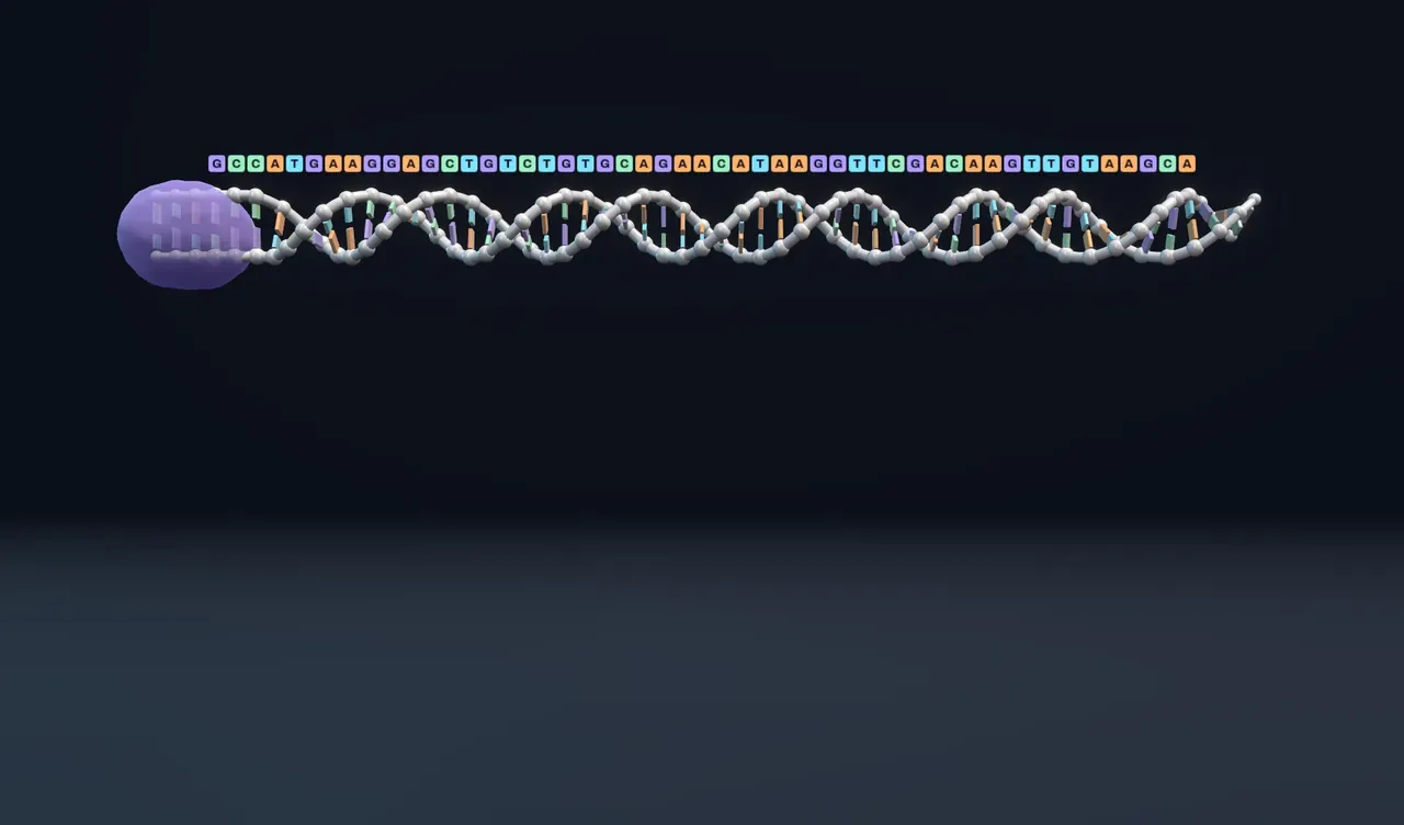
DNA stays put. In cells like ours it never leaves the nucleus, so the cell works from a copy. A machine called RNA polymerase settles on the DNA just before the gene, prises the two strands a little way apart and runs along one of them. As it goes it builds a single strand of messenger RNA, matching each letter as the strands pair.
The copy carries the same message as the gene with one swap: RNA uses U (uracil) where DNA uses T. This first step is called transcription.
Try it: drag Make the protein a little way from Gene and follow the polymerase. Each letter of RNA lands directly below the DNA letter it copies, with every T turned into a U.
Step two: read it three letters at a time
The RNA travels to a ribosome, a machine of RNA and protein that builds the chain. The ribosome slides along the RNA until it finds the letters AUG, the start signal, and from there it reads the message in groups of three. Each group is a codon, and each codon stands for one amino acid. This step is translation.
Four letters taken three at a time make sixty-four codons. Sixty-one of them name an amino acid, so most amino acids have several codons; the last letter of a codon often makes no difference. The other three codons, UAA, UAG and UGA, mean stop. The table that links codons to amino acids is the genetic code, and with a handful of small exceptions every living thing uses the same one.
The ribosome cannot recognise an amino acid by itself. For each codon a small adapter molecule called transfer RNA (tRNA) arrives carrying the right amino acid. Three letters at its tip pair with the codon, so only the matching tRNA fits. The ribosome joins its amino acid onto the growing chain, the empty tRNA leaves, and the ribosome steps on three letters. A bacterial ribosome adds around fifteen amino acids a second.
Step three: the chain folds
A freshly made chain is a floppy string, and a string cannot catch, cut or carry anything. Its job depends on its shape. Some amino acids are oily: they are mostly carbon and hydrogen and, like oil in water, they avoid the water around them. Others are water-loving, carrying electric charges or groups that water molecules cling to. As the chain jostles about, the oily ones end up packed together in the middle and the water-loving ones on the surface.
Because the order of oily and water-loving amino acids differs from protein to protein, each folds into its own shape. Real folding also depends on hydrogen bonds, spiral and sheet-like stretches and many other details, and some proteins need helper proteins to fold at all, but the oily core is the main reason a chain folds into a ball rather than staying loose.
Try it: with Make the protein at Folded protein, look at the middle of the fold. In the original protein all four central places hold oily amino acids, and the lit links show oily neighbours touching.
One letter changed
A change in a gene’s letters is a mutation. The simplest kind swaps, adds or removes a single letter, and the experiment above shows the four things that can follow.
- Silent. The new codon names the same amino acid. CTG and CTA both mean leucine, so the protein is unchanged.
- Missense. The new codon names a different amino acid. Here GTG (valine, oily) becomes GAG (glutamate, water-loving). An oily amino acid that sat in the core now wants to face the water, and the chain folds another way. The real-life example is sickle-cell disease: one letter in the gene for part of haemoglobin, the oxygen carrier in red blood cells, turns a GAG into GTG, the reverse of the change here. The protein still folds, but an oily patch on its surface makes the molecules stick together into stiff fibres.
- Nonsense. The new codon is a stop. CAG becomes TAG, and the ribosome lets go after six amino acids. A protein cut that short usually cannot work at all.
- Frameshift. One letter is lost, or one extra is slipped in. The ribosome still reads in threes from the start, so every codon after the change is grouped wrongly, the amino acids come out as nonsense, and a stop codon usually turns up by chance soon after.
Try it: choose Frameshift and watch the codon brackets under the RNA. Then choose Missense and compare the new fold with the faded original beside it.
Losing three letters together is different from losing one: the reading frame survives, and the protein simply misses one amino acid. The most common mutation behind cystic fibrosis is exactly that, three letters lost and one amino acid gone.
What this model leaves out
The gene is made up and very short: fourteen amino acids and a stop codon, where a typical human protein has a few hundred amino acids. In cells like ours the RNA copy is also trimmed and spliced before it leaves the nucleus, and many ribosomes read the same RNA at once. None of that is shown.
The fold is computed, not drawn by hand, but by a deliberately simple rule. Each amino acid is treated as either oily or water-loving and placed on a flat square grid, and the fold shown is the one that brings the most oily pairs side by side; among equally good folds the most compact wins. This is the HP model, which scientists use to study why proteins fold. It captures the oily core and nothing else. Real proteins fold in three dimensions, and predicting their exact shapes takes far more detailed physics or computer models trained on thousands of known structures.
Sizes are not to scale. RNA polymerase and the ribosome are drawn small so that the letters stay readable; a real ribosome is about as wide as thirty letters of RNA, and the amino acids are drawn as beads spaced to sit under their codons.