A shadow stacks everything
A plain X-ray picture is a shadow. X-rays pass through the body to a detector, and wherever they cross bone more of them are stopped, so the bone shows white. But each point of the picture records everything along one straight line through the body: ribs, heart, spine and lungs are piled on top of one another. A shadow cannot tell you how deep anything is.
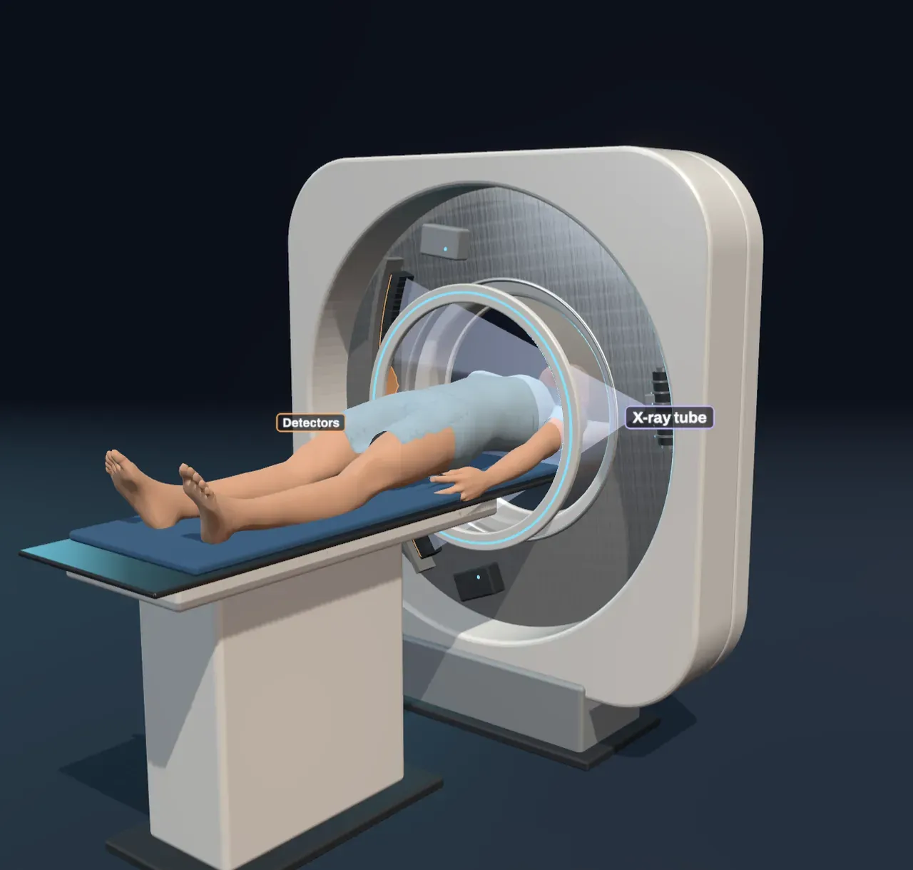
A CT scanner (computed tomography: drawing by slices) gets round this. Its X-ray tube and a curved row of detectors sit on opposite sides of a ring, the gantry, and spin round the patient two to four times a second. The table slides the body through the ring, and each turn measures one thin slice.
Every reading is a sum along a line
Each detector reading says how strongly the X-rays were dimmed on their way from the tube. Every tissue weakens the beam by a fixed fraction per centimetre, its attenuation. Bone weakens it most, water-rich muscle and organs less, fat a little less again, and the air in the lungs hardly at all. Over a whole path the fractions multiply, so the logarithm of the dimming is simply the sum of the attenuation along the line.
One sweep across the detector, with the tube at one angle, gives a projection: a profile of those sums. Stack the profiles from every angle, one row each, and you get a sinogram. Each point inside the body traces a sine-shaped stripe through it, because its shadow slides from side to side as the tube goes round.
From shadows back to a slice
How do you undo the sums? Take each profile and smear it back across the image along the lines it was measured on. Where many smears from different angles agree, something dense must be there. This back-projection alone gives a blurred image, so each profile is first sharpened by a filter that boosts fine detail and adds a slight dip beside every edge. Summed over every angle, the dips cancel the blur exactly. This filtered back-projection is what the scanner’s computer does, and the mathematics behind it was published by Johann Radon in 1917, long before anyone could use it. Godfrey Hounsfield built the first clinical scanner in 1971.
Try it: choose Head under Slice position, set Projection angles to 6 and choose Slice under Build the slice. Then slide it up to 180 and watch the streaks fade.
How many angles?
With only a few views, every sharp edge smears into straight streaks that point back towards the angles that were measured, and the skull or the spine throws spokes across the whole slice. As views are added, the streaks from different directions overlap and cancel, and the real shapes emerge: the black lungs, the bright vertebra, the grey liver and kidneys. A real scanner takes about a thousand views every turn.
The slider also shows why the method cares about dose. Each view needs X-rays, and X-rays can damage cells. A chest CT gives a dose of several millisieverts: about two years of natural background radiation, and a few hundred times a chest X-ray. Scanners are designed to get a clear slice from as few X-rays as they can.
Other ways in
CT shows bone, air and dense tissue sharply, but soft tissues that attenuate X-rays almost equally look nearly the same grey. MRI instead measures radio signals from hydrogen nuclei in a strong magnet, and it tells soft tissues apart with no ionising radiation. Ultrasound times echoes of high-pitched sound bouncing off boundaries inside the body; it is cheap, safe and moving, but it cannot see through bone or gas. Doctors choose the tool by what they need to see.
What this model leaves out
The patient’s insides are modelled as a phantom: ellipses with fixed attenuation values, and its organs carry the brightening of an injected contrast agent, so they stand out a little more than they would without one. The scene computes each view exactly, with no photon noise, scattered X-rays or beam hardening (the low-energy part of the beam being soaked up first), all of which add grain and artefacts to real images. Real scanners measure many slices at once with dozens of detector rows, often while the table moves in a spiral, and use far finer grids than 128 × 128. The drum is slowed down enormously, and the X-ray fan, invisible in reality, is drawn with its brightness compressed so the shadow of the body shows.