Why cells divide
You began as a single cell, a fertilised egg. Everything you are now, tens of trillions of cells, came from that one cell dividing, and its daughters dividing, over and over. Division never stops: skin flakes away and the lining of your gut wears out in days, so your body replaces roughly a third of a trillion cells every day: several million every second.
Each new cell needs its own complete copy of the instructions, the DNA. So the hard part of division is not splitting a bag of chemistry in two. It is making sure each half gets exactly one copy of every chromosome, no more and no fewer.
Copy first, then sort
Most of a cell’s life is spent in interphase, growing and doing its job. Before it divides, it copies all its DNA. Each chromosome now consists of two identical strands, called sister chromatids, held together at a pinched point, the centromere. At this stage the DNA is spread out as fine threads, far too tangled to sort.
Division then sorts the copies in a fixed order, the stages of mitosis, a word from the Greek for thread, coined by Walther Flemming in 1882 after watching the threads in stained cells.
Four stages and a split
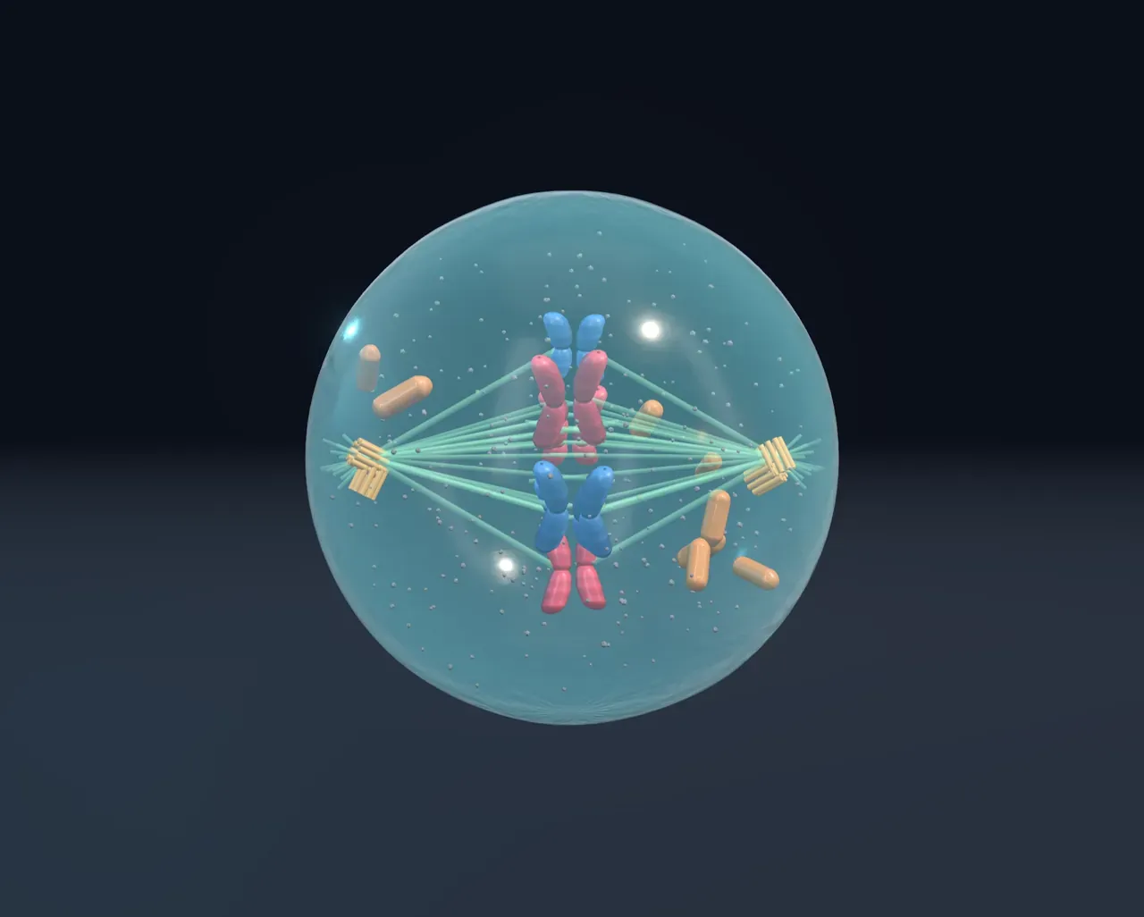
In prophase each chromosome winds itself up tightly into a short, thick X: the two sisters side by side. The nuclear envelope breaks into pieces, and two centrosomes move to opposite sides of the cell, growing a spindle of protein fibres between them. In metaphase fibres from both sides grip every chromosome at its centromere and tug it into a line across the middle. In anaphase the protein rings holding the sisters together are cut, and the fibres reel each sister towards opposite poles. In telophase a new nucleus forms around each complete set. Finally, in cytokinesis, a ring of contracting protein tightens round the cell’s waist until it pinches into two.
Try it: press Metaphase, then choose On under Show the spindle. Now press Anaphase and watch each X split into two.
A full set for each daughter
Chromosomes come in pairs: one of each pair came from your mother, the other from your father. In the scene they are pink and blue. Humans have 46 chromosomes in 23 pairs; a fruit fly has 8 and a pea plant 14. Change Chromosome pairs and notice that, whatever the number, each daughter cell ends up with one copy of every chromosome, pink and blue alike. Mitosis makes two cells genetically identical to the original.
Eggs and sperm are made by a different division, meiosis. It divides twice after a single copy, so it makes four cells with one chromosome from each pair: 23 in a human egg or sperm. When they meet, the number is 46 again.
When division goes wrong
A healthy cell divides only when its neighbours signal that more cells are needed, and it checks itself along the way: at several checkpoints the cycle halts if the DNA is damaged or a chromosome is not attached to the spindle. Cancer begins when mutations break those controls. The cell divides when it should not, ignores damage, and passes the faults to its daughters, which can grow into a tumour. Many cancer drugs work by jamming the spindle.
What this model leaves out
The scene shows a handful of chromosomes, while a human cell sorts 46, each of them thousands of times longer when unwound than it appears here. Real cells are crowded with far more organelles, and the two daughters stay joined by a thin bridge for a while before it is cut. The pink and blue colours are a teaching device. The timing is uneven too: in a typical human cell growing in a dish, the whole cycle takes about a day, and mitosis only an hour of it.