From air to nerve
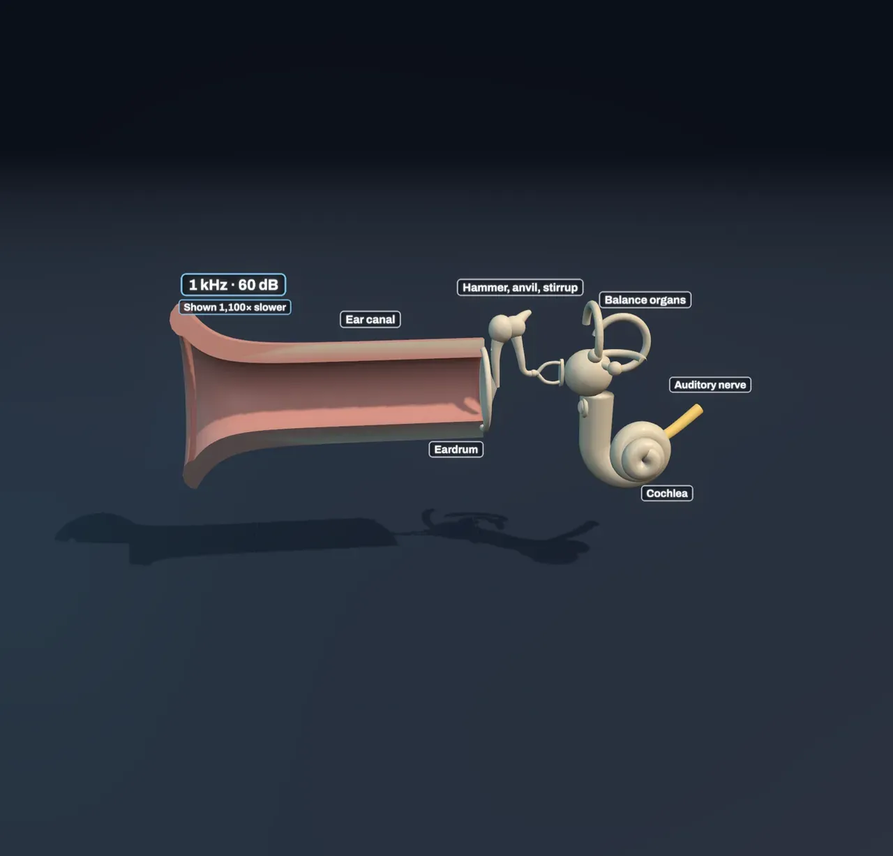
Sound is a travelling squeeze and stretch of the air. When it reaches you, it runs down the ear canal, a tube about 2.5 cm long, and pushes on the eardrum at its end: a thin, taut membrane a little under a centimetre across. The eardrum moves in and out with every squeeze, and a chain of three tiny bones carries the movement across the air-filled middle ear to the inner ear, buried in the hardest bone of the skull.
There the movement enters the cochlea, a coiled tube of fluid about the size of a pea. Inside it, the sound is turned into signals in the auditory nerve, and the brain hears it. The scene shows all of this at its true size, with the canal cut open so you can see in.
Why the middle ear has bones
Sound in air barely gets into water: almost all of it bounces off the surface. The fluid in the cochlea has the same problem, and the middle ear is the fix.
The eardrum is about seventeen times larger in area than the footplate of the stirrup, the last bone, which sits in a small opening called the oval window. The same push gathered over the big eardrum and delivered through the small footplate becomes a much bigger pressure. The hammer and anvil add a little lever action on top, so the pressure on the fluid ends up more than twenty times what reached the eardrum.
Try it: watch the bones while you change Loudness. The hammer’s handle is fixed to the eardrum; the hammer and anvil rock together about their heads, and the stirrup moves in and out of the oval window like a piston.
A wave that sorts pitch
Running the length of the cochlea, dividing it in two, is the basilar membrane. It is narrow and stiff at the base, near the oval window, and gets wider and floppier towards the tip, about 3.5 cm along. Each part of it would rather vibrate at its own pitch: the stiff base at high pitches, the floppy tip at low ones.
When the stirrup pushes on the fluid, a wave starts along the membrane at the base and travels towards the tip. As it goes, it slows and grows, until it reaches the place that suits its pitch. There it peaks, and within a millimetre or two it dies away. A high pitch peaks near the base; a low pitch travels almost to the tip. So the cochlea sorts sound by pitch along its length, with each doubling of pitch moving the peak a few millimetres.
Try it: choose Cochlea uncoiled under Uncoil the cochlea, so the cochlea uncoils into a straight tube. Then move Pitch (powers of ten) from Low hum 50 Hz to Mosquito tone 17 kHz and watch the peak slide from the tip to the base.
The slider uses powers of ten because the membrane is laid out much the same way: equal ratios of pitch take up roughly equal lengths. From 100 Hz to 1,000 Hz covers about 11 mm of it, and from 1,000 Hz to 10,000 Hz about 16 mm. Georg von Békésy first watched these waves in cochleas from cadavers, and won a Nobel Prize for it in 1961.
Hair cells turn motion into signals
Sitting on the membrane in a row along its whole length are about 3,500 inner hair cells. Each has a tuft of stiff bristles on top. When the membrane moves up, the bristles bend, tiny channels in them open, and the cell passes a signal to the nerve fibres touching it. About 30,000 fibres leave the cochlea in the auditory nerve, and most of them listen to one inner hair cell each.
Because each hair cell sits at one place on the membrane, each fibre stands for one pitch. The brain learns which pitch it is hearing from which fibres are busy. A chord, or a voice, sets several places moving at once, and the brain hears them all.
About 12,000 outer hair cells do a different job: they push back on the membrane in time with it, making quiet sounds bigger and the peak sharper. The scene leaves them out.
Loud and quiet
Loudness is measured in decibels. Each 10 dB is ten times more sound energy: a quiet room is about 30 dB, a conversation about 60, a busy street 80, and a rock concert over 100. A louder tone moves the membrane further, so each fibre fires faster, and the vibration spreads to more places, mostly towards the base, so more fibres join in.
The quietest tone you can hear depends on its pitch. Ears are most sensitive between about 2 and 5 kHz, partly because the ear canal resonates near 3 kHz, and much less sensitive at the ends of the range. A 50 Hz hum must be over 40 dB before a young adult hears it at all.
Try it: choose Low hum 50 Hz and Whisper. The wave reaches the tip, but no hair cell moves enough to fire. Raise Loudness until the nerve lights up.
Why high pitches fade first
Every sound, whatever its pitch, passes the base of the cochlea on its way along the membrane. Over a lifetime the hair cells there take the most wear, and hair cells that die are not replaced. That is why hearing usually fades from the top down: by 70, most people have lost the highest pitches altogether, and hear 4 to 8 kHz much less well. Loud noise does the same damage faster, and 85 dB for hours a day is enough to do it.
Try it: choose Mosquito tone 17 kHz and Busy street, then change Listener’s age from 20 years to 45 years. The same tone is no longer heard. Uncoil the cochlea and the base of the membrane is missing many of its hair cells.
What this model leaves out
The scene slows every vibration by a factor shown above the canal, from about 40 times for a 20 Hz tone to more than 13,000 for 20 kHz, and it magnifies every movement: a whisper moves the eardrum less than the width of an atom. The amplitudes are also compressed, so that quiet and loud tones both show.
The shape of the wave along the membrane, the way firing spreads with loudness and the hair-cell losses are simple fits to measured behaviour, not a simulation of the cochlea’s fluid. The real cochlea is divided into three fluid-filled channels, and the membrane carries a whole structure, the organ of Corti, rather than a single row of cells. Nerve fibres also fire in step with the wave for low pitches, which gives the brain a second clue to pitch. Two muscles in the middle ear, which stiffen the bones against loud sounds, and everything the brain does with the signals from both ears, are not shown.