Chemistry at body temperature
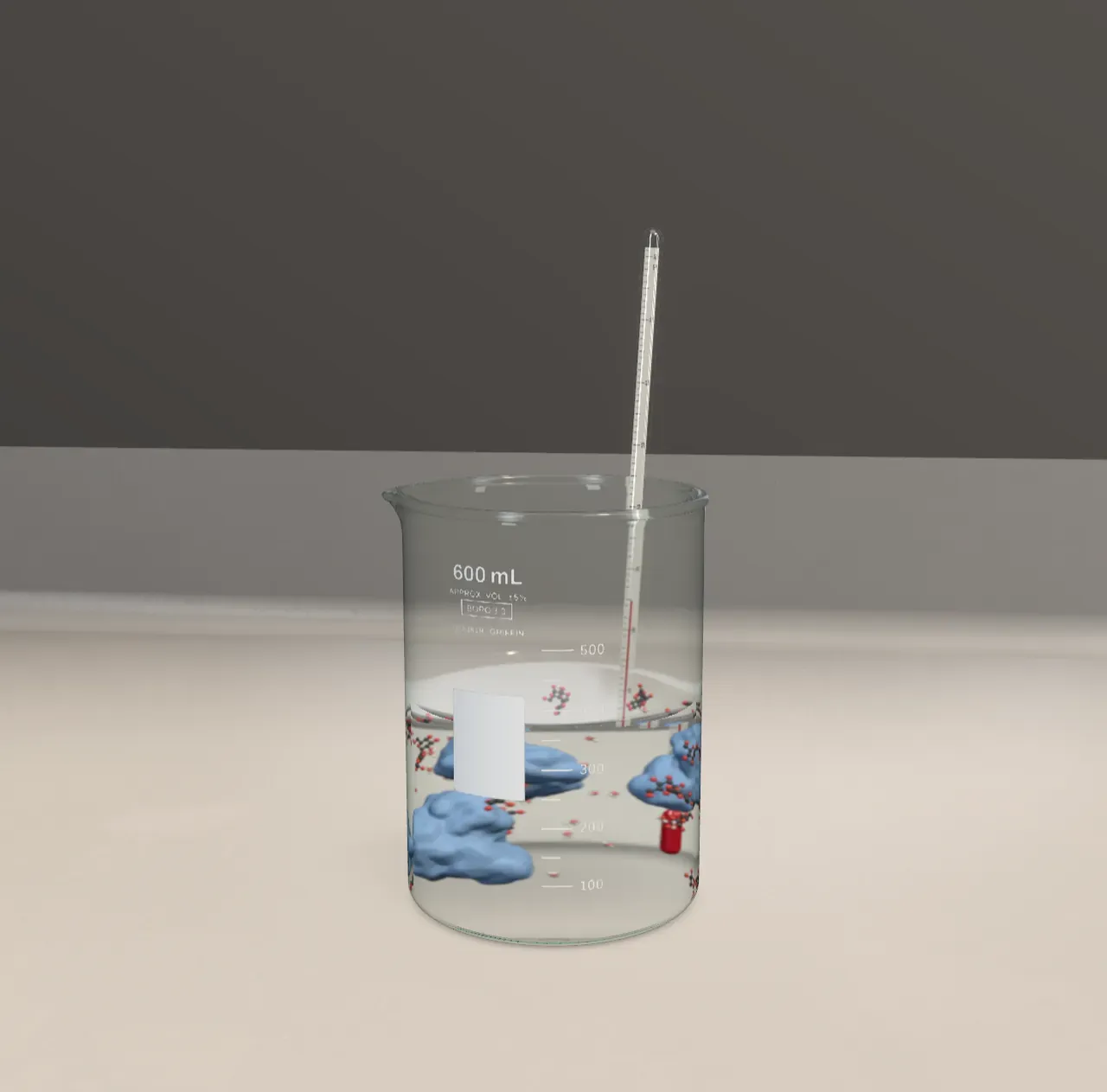
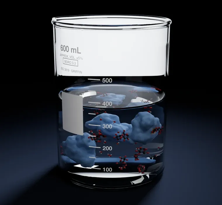
Saliva contains an enzyme, amylase, that starts breaking starch into sugar while you chew, which is why bread held in the mouth for a minute begins to taste sweet. Further along the gut, other enzymes split those sugars into single glucose molecules that the body can absorb. The beaker shows that last step.
Sugar and water left alone would take an extremely long time to react at 37 °C; the activation energy is too high. Cells cannot simply heat themselves up to speed things along, so they use enzymes: proteins that act as catalysts. Many speed their reaction by a factor of millions or more. Catalase, which destroys the hydrogen peroxide that cells make, can handle millions of molecules every second.
A pocket with the right shape
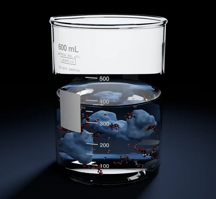
An enzyme is a long chain of amino acids that folds into a precise three-dimensional shape. Somewhere on its surface is a cleft called the active site. Only molecules of the right shape and charge fit there. In 1894 the chemist Emil Fischer compared this to a lock and key. Later, Daniel Koshland proposed that the fit is more like a hand slipping into a glove: the site shifts a little to close around its target, an idea called induced fit.
Once held in the site, the sugar is strained and its bonds are pulled at just the right angles, giving the reaction an easier path with a lower barrier. The two halves are released, the site is empty again, and the enzyme takes the next molecule.
Warmer is faster, until it isn’t
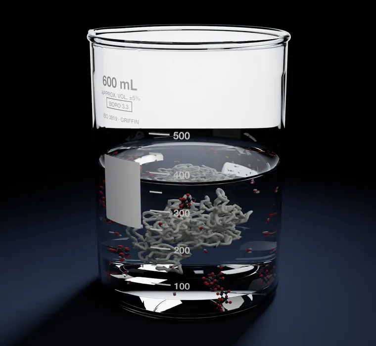
Warm the beaker and the molecules jostle harder. More arrive at the active sites, and more of them carry enough energy to react, so the rate climbs. But the enzyme’s fold is held together by many weak attractions between parts of the chain. A few degrees above body temperature those start to shake loose, the chain unravels, and the active site loses its shape. The enzyme is denatured, and the rate collapses. Cooking an egg does the same to its proteins: the clear white turns solid and opaque.
Try it: choose Fridge, then Body, then Hot bath, and watch each folded enzyme loosen into a bare chain. Watch the activity curve as you go: the rate rises and then falls. Choose On under Fade the water to see the enzymes and sugars more clearly.
pH matters too
The active site’s grip depends on electric charges, and the pH changes those charges. Most enzymes have a pH at which they work best. Pepsin, which digests protein in the acidic stomach, works best around pH 2; trypsin, which carries on the job in the small intestine, prefers about pH 8. Move the pH control away from 7 and fewer sugars stay in the site long enough to be split.
What this model leaves out
Each enzyme is the real fold of human maltase, from its crystal structure, but drawn as a smooth surface: the thousands of atoms that hold the sugar with many precise contacts are not shown, and the sugars are drawn about four times too large so their atoms can be seen. An unfolded enzyme is drawn as its bare backbone, one of a few loose shapes rather than a real tangle. The model unfolds each enzyme over a narrow band of temperatures and refolds it when cooled; a real enzyme heated too far usually tangles and stays useless. The temperature curve is a typical shape for an enzyme from the human body, while enzymes from microbes in hot springs keep working near boiling.