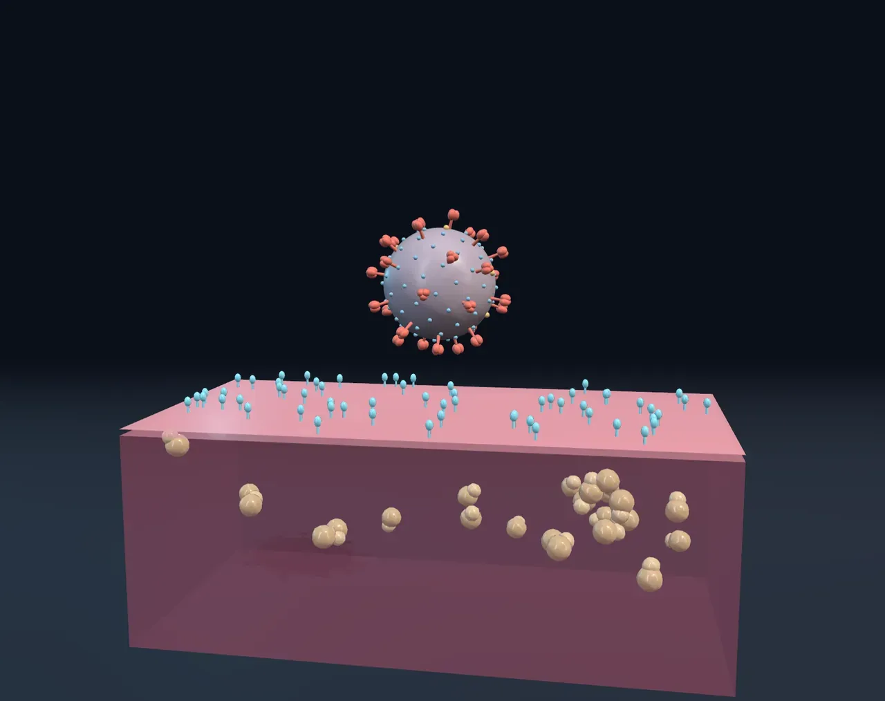
Smaller than anything alive
In the 1890s, researchers studying a disease of tobacco plants found that sap stayed infectious even after passing through filters fine enough to catch every bacterium. Whatever caused it was smaller than any known germ. It was a virus, and nobody actually saw one until electron microscopes arrived in the 1930s.
Most viruses measure between about 20 and 300 nanometres, millionths of a millimetre. Line up twenty coronaviruses and they would just span one E. coli bacterium; it takes about ten of those bacteria to span a typical human cell. Ordinary light microscopes cannot make out anything much smaller than 200 nanometres, so most viruses are simply invisible to them.
Genes in a box
A virus particle is little more than instructions in packaging. At its centre is a genome: a single long molecule of DNA or RNA, or for influenza eight separate pieces. Around it is a capsid, a shell built from many copies of the same few proteins, often fitted together into a twenty-sided shape. Some viruses, such as coronaviruses and influenza, also wear an envelope: a scrap of fatty membrane taken from the last cell they left, studded with their own proteins.
What a virus lacks matters as much. It has no machinery to make energy, no way to build proteins, and no means of copying itself. Outside a cell it is inert, which is why biologists still argue over whether to call viruses alive.
Breaking in
A virus drifts until it bumps into a cell carrying the right molecule on its surface. The fit is specific: the coronavirus behind COVID-19 grabs a protein called ACE2, influenza grabs sugars on the cells lining your airways, and the bacteriophage T4 grabs the outer coat of the gut bacterium E. coli. That fit decides which hosts, and which tissues, a virus can infect.
Then the genes have to get in. Many animal viruses are swallowed whole in a pocket of membrane and break open inside. T4 does something more mechanical: it lands on six long legs, contracts the spring-loaded sheath around its tail, drives a hollow tube through the cell’s envelope and pumps its DNA through it.
Try it: choose Phage T4 and drag Infection slowly from Attached to Inside. Watch the tail sheath shorten as the DNA leaves the head. Then choose Cut open under Cut open on a Coronavirus.
A factory taken over
Once inside, the viral genes are read by the cell’s own machinery as if they were the cell’s own. The cell starts copying the viral genome and building viral proteins, and the new parts assemble themselves into fresh particles. A T4 infection can turn one bacterium into a hundred or more new phages in about half an hour. The new viruses then leave, either budding out wrapped in a piece of membrane or bursting the cell open.
Why antibiotics don’t work
Antibiotics attack structures that only bacteria have, such as their cell walls and their own kind of protein-building ribosomes. Viruses have neither, so antibiotics cannot touch them; a cold or flu is caused by a virus, so antibiotics do not help. Some antiviral drugs exist, aimed at steps in a particular virus’s cycle. Vaccines work differently: they show the immune system a harmless version, piece or blueprint of a virus, so it is ready to recognise the real thing.
What this model leaves out
The Scene follows one particle and shows four offspring. Real cells are far larger than the patch shown, and real infections make hundreds to thousands of copies. Coronaviruses actually assemble on membranes deep inside the cell rather than budding at its surface, and adenovirus copies its DNA inside the cell’s nucleus, which is not drawn.