A folded sheet of neurons with a map of jobs: seeing at the back, movement in a strip across the top, crossed wiring so each side of the brain serves the opposite side of the body, speech that lives on the left, and neurons that fire faster when their area works.
Step 1 of 6 · A folded sheet
Step 1 of 6
A folded sheet
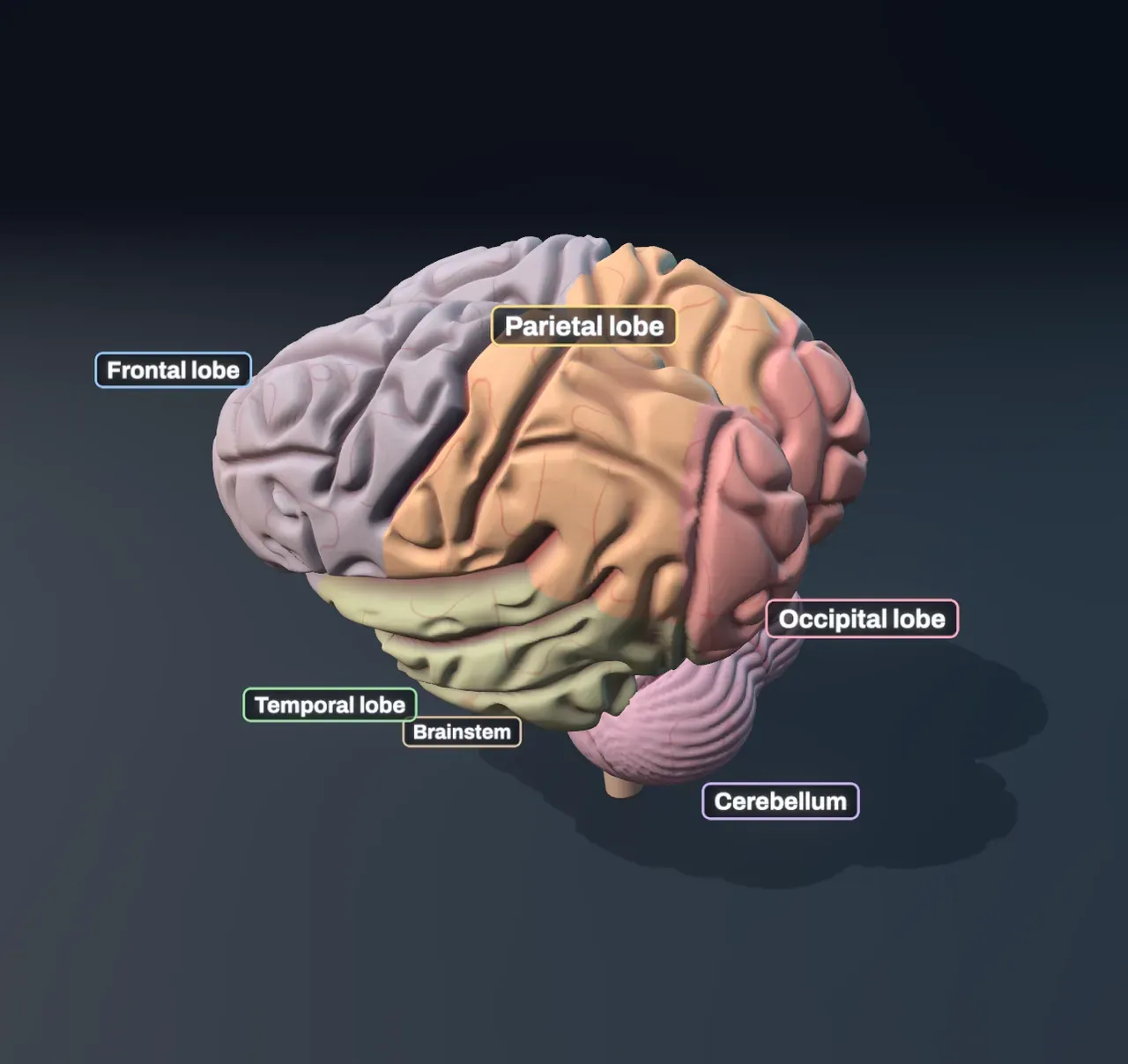
Most of what you see is the cerebrum, split into two hemispheres. Its surface is the cortex, a sheet of neurons two to four millimetres thick, folded so it fits in the skull. Deep grooves split each hemisphere into four lobes: frontal, parietal, temporal and occipital. Underneath, at the back, is the cerebellum, which times movements, and the brainstem joins it all to the spinal cord. Zoom to the neurons is at Lobes.
Step 2 of 6
Crossed wiring
Zoom to the neurons is at Active areas: the brain turns grey, and the areas the task uses glow in brain-scan colours. The task is Moving a hand, the left hand, and the glow is in the motor cortex, a strip across the top of the brain just in front of the central groove.
Step 3 of 6
A busy patch
Zoom to the neurons is at Neurons: a patch of the visual cortex in the left hemisphere. Every neuron speaks in impulses, brief all-or-nothing pulses. The chart shows one second in the patch as a raster: a row for each neuron, a line for each impulse, and the task’s neurons at the top. With nothing to do, a typical cortex neuron fires about once a second. For now, the thing you are looking at is on your left.
Step 4 of 6
Speech lives on the left
Speaking uses Broca’s area, in the frontal lobe, and the mouth part of the motor strip. In about 95% of right-handers, speaking and understanding speech rely mostly on the left hemisphere. The chart shows what share of the extra activity is on the left.
Step 5 of 6
What a scan sees
The glow in the Scene is like an fMRI scan: a map of a difference, the brain doing a task minus the same brain at rest. But the scanner does not see the impulses themselves. Switch Task between Hearing, Seeing and Moving a hand, and see how many areas work for each.
Step 6 of 6
Never idle
Choose Resting: a different set of areas glows, in both hemispheres, and the seeing and moving areas fade. This is the default mode network, which works while the mind wanders. The brain weighs about a kilogram and a half, about 2% of the body, yet at rest it uses about a fifth of the body’s energy.
Loading the interactive version…
Loading the interactive version…
Each step sets up the Scene for you, and a control marked “fixed” stays put for that step.
Practice · Choose
In which lobe is the visual cortex, where seeing starts in the brain?
Practice · Choose
In which lobe is the visual cortex, where seeing starts in the brain?
Practice · Predict, then look
Moving the left hand lights the motor strip of the right hemisphere. Predict what glows when Side switches to Right.
Practice · Predict, then look
Moving the left hand lights the motor strip of the right hemisphere. Predict what glows when Side switches to Right.
Practice · Predict, then look
The patch in the left visual cortex is almost silent, because what is on your left is seen by the right hemisphere. A cortex neuron with nothing to do fires about once a second. Predict how many times a second, on average, the task’s neurons fire when Side switches to Right.
Practice · Predict, then look
The patch in the left visual cortex is almost silent, because what is on your left is seen by the right hemisphere. A cortex neuron with nothing to do fires about once a second. Predict how many times a second, on average, the task’s neurons fire when Side switches to Right.
Practice · Predict, then look
While speaking, about 62% of the extra activity is in the left hemisphere. Predict what happens when Side switches to Left.
Practice · Predict, then look
While speaking, about 62% of the extra activity is in the left hemisphere. Predict what happens when Side switches to Left.
Practice · Choose
What does an fMRI scanner really measure when it shows a coloured patch in the hearing cortex?
Practice · Choose
What does an fMRI scanner really measure when it shows a coloured patch in the hearing cortex?
Practice · Choose
At rest, with the mind wandering, most of the seeing and moving areas fade. Does that mean that most of the time we use only a small part of the brain, such as “10%”?
Practice · Choose
At rest, with the mind wandering, most of the seeing and moving areas fade. Does that mean that most of the time we use only a small part of the brain, such as “10%”?
Lesson complete. Every practice question here is done.