A folded sheet
An adult brain weighs about 1.4 kilograms, some 2% of the body, yet at rest it uses about a fifth of the body’s energy: roughly 20 watts, the power of a dim light bulb. It is soft, about as firm as tofu, and floats in a cushion of fluid inside the skull.
Most of what you see from outside is the cerebrum, split down the middle into two halves, the left and right hemispheres. Its surface is the cortex, a sheet of nerve cells only 2 to 4 millimetres thick. Spread flat, the cortex of both halves would cover about 0.2 square metres, a square 45 centimetres on a side. Folding is how that much sheet fits inside a skull: about two-thirds of it is hidden in the grooves. Each ridge is a gyrus and each groove a sulcus. Below the grey sheet lies the white matter, the wiring: bundles of nerve fibres that join one part of the cortex to another and to the rest of the body.
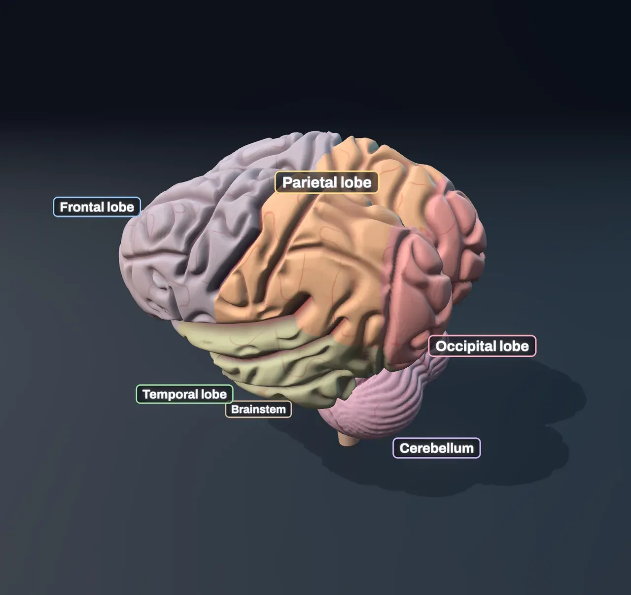
A few deep grooves divide each hemisphere into four lobes: the frontal lobe behind the forehead, the parietal lobe under the crown, the temporal lobe above the ear and the occipital lobe at the back. Tucked underneath at the back is the cerebellum, the “little brain”, which smooths and times movement. It holds about 69 of the brain’s 86 billion neurons, packed into its fine folds. The brainstem joins it all to the spinal cord and runs the things you never think about, such as breathing and the heartbeat.
Try it: with Zoom to the neurons at Lobes, drag the brain round to see the gap between the two halves from above, and the cerebellum’s fine folds from below.
A map of jobs
The cortex is not the same everywhere. Different patches do different jobs, and the map is much the same from one person to the next.
The first clues came from injuries. In 1861 the French doctor Paul Broca examined a man who understood what was said to him but could say only one syllable, “tan”. After the man died, Broca found damage in the left frontal lobe, just above the temporal lobe: the place now called Broca’s area. In 1874 Carl Wernicke described patients with damage further back, in the left temporal lobe, who spoke fluently but made little sense and could not follow what others said. That place is Wernicke’s area.
In the 1930s and 1940s the surgeon Wilder Penfield mapped the cortex of patients who were awake during brain surgery for epilepsy (the brain itself feels no pain). A small current on one strip, just in front of the central sulcus, made a finger or a lip twitch: the motor cortex. On the strip just behind it, the current made patients feel a touch on their skin: the touch cortex. Each strip holds a map of the body, upside down, with the feet at the top and the face at the bottom, and with huge shares for the hands, lips and tongue, which do the finest work.
Seeing starts in the visual cortex at the very back of the brain, as far from the eyes as it can be. From there, signals flow forwards along two routes: down into the temporal lobe to work out what things are, and up into the parietal lobe to work out where they are and how to reach them. Sounds arrive at the hearing cortex, tucked into the deep groove above the temporal lobe. How the eye and the ear turn light and sound into signals is told in vision and hearing.
Try it: step Zoom to the neurons to Active areas and click through Task. Watch the glow move from the back of the brain (Seeing) to the temporal lobe (Hearing) to the strip across the top (Moving a hand).
Crossed wiring
Most of the wiring between the cortex and the body crosses over. Each half of the brain moves and feels the opposite side of the body. The fibres that carry commands from the left motor cortex cross to the right side low in the brainstem and run down the spinal cord to the muscles of the right hand. A stroke in the left half can paralyse the right side of the body.
Sight crosses in its own way. Each half of the brain sees the opposite half of the view, through both eyes: the nerve fibres from each eye split where the two optic nerves meet, so everything to your right ends up in the left visual cortex. Hearing crosses only partly. Each ear sends signals to both halves, the opposite one a little more strongly.
The cerebellum is the exception. It works with its own side of the body, so moving your right hand uses the left motor cortex and the right half of the cerebellum.
Try it: choose Moving a hand and switch Side between Right and Left. The glow jumps from the top of the left half to the top of the right half, and the cerebellum’s glow swaps sides the other way.
No task uses just one spot
A brain scan in a hospital or lab, called functional MRI, does not see neurons fire. It sees blood. Working neurons need more oxygen, and within a few seconds the blood flow to a busy patch rises by more than they use, so the blood there carries more oxygen. The scanner picks up that difference. The colourful blobs in pictures of scans, like the glow in the Scene, are a map of a difference: the brain doing a task, minus the same brain resting. The changes are small, a few per cent, and the brain’s total energy use barely rises when you think hard.
So the idea that we use only 10% of our brains is a myth. Every part is active all the time; a task makes some parts busier than they were. And no task uses just one spot. Seeing an object lights the visual cortex and areas forward of it; speaking needs Broca’s area, the mouth part of the motor strip, areas that plan the movement, the hearing cortex to hear your own voice and the cerebellum to keep the timing. The areas work as a network, passing signals to each other along the white matter.
Some areas are busier at rest than during a task. When you lie still with nothing to do and your mind wanders, to memories, plans or other people, a set of areas along the middle and sides of both halves becomes more active, and it quietens again as soon as you turn to a task. It is called the default network.
Try it: choose Resting. A different set of areas glows, in both halves, and the visual and motor areas fade.
Inside a busy patch
Step Zoom to the neurons to Neurons and the Scene zooms into a patch of the left half’s cortex, in the area the task leans on most. The cortex is built in six layers, numbered from the surface down. Signals from the senses arrive in the middle, in layer IV, along fibres from the thalamus, a relay station deep in the brain. Big pyramidal neurons in layer V send their long fibres down into the white matter and on to other areas, the brainstem or the spinal cord. Smaller interneurons connect cells close by, mostly to calm them down.
Each neuron talks in spikes: the brief all-or-nothing pulses described in the nervous system. A busy patch is one where many neurons spike faster. With nothing to do, a typical cortical neuron fires about once a second. When its area works, the neurons the task uses fire tens of times a second, while their neighbours barely change.
The chart under One second in the patch shows this as a raster: one row per neuron, one tick per spike, the neurons the task uses at the top. The pattern, which cells fire and when, carries the information. Artificial neural networks borrowed a much simplified version of this idea.
Try it: with Zoom to the neurons at Neurons, choose Seeing, then switch Side to Left. The patch in the left visual cortex goes quiet: what is on your left is seen by the right half.
Speech lives on the left
Language is lopsided. In about 95% of right-handed people, and in about 70% of left-handed people, speaking and understanding speech depend mostly on the left half. The right half still helps, with the melody and emotion of speech, and the two halves share nearly everything else.
That is why the Side choice changes nothing for Speaking: Broca’s and Wernicke’s areas stay on the left. The motor strips of both halves work, because both sides of the lips and tongue move.
Try it: choose Hearing and switch Side. The hearing cortex on the side opposite the ear works harder, but Wernicke’s area, which makes sense of the words, stays on the left.
What this model leaves out
The activity levels are made up to follow the textbook pattern, not measured in one person. Real scans differ from person to person and from moment to moment, and their blobs have ragged edges and spread over many more small patches. A real task uses deep structures too, such as the thalamus and the basal ganglia, which the Scene does not show, and its activity changes over time: a signal from the eyes reaches the visual cortex in about a twentieth of a second, and the areas further forward a little later.
Each area is drawn where it usually sits, on a brain whose folds are generated by the computer. Real brains share their big grooves, but the small folds differ from person to person and even between the two halves. Much of the visual and hearing cortex lies hidden inside grooves or on the inner face of each half, so the glow on the surface shows only part of it.
The patch shows a few dozen neurons where a real piece of cortex this size has hundreds of thousands, each with thousands of connections rather than three. Its spikes are random at the rate the task sets for each cell, slowed twenty times; in a real brain the timing between cells matters, and some patterns come in waves. The senses of touch, taste and smell, and how the brain combines them, are left to a later Article on the senses.