Two pumps side by side
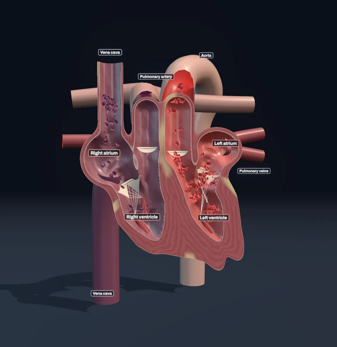
Put your fingers on the inside of your wrist and you can count it: about seventy times a minute, a hundred thousand times a day, a fist-sized muscle squeezes and lets go. Every minute it moves about 5 litres of blood, which is all the blood you have. The Scene shows a heart cut in half from the front, as if you were facing the person whose heart it is, so its right side is on your left.
The heart is two pumps joined by a shared wall. The right side takes in blood that has been round the body and is low in oxygen, and pumps it to the lungs. The left side takes in blood coming back from the lungs, rich in oxygen, and pumps it round the body. Each side has two chambers: a thin-walled atrium on top that collects blood as it arrives, and a muscular ventricle below that does the pumping.
Look at the cut walls. The left ventricle’s is about a centimetre thick, three times the right’s, because it has to drive blood through every organ from your scalp to your toes, while the right ventricle only has to push it through the lungs next door.
The Scene colours the blood by how much oxygen it carries: plum on the right side, scarlet on the left. Real blood low in oxygen is dark red, never blue. Veins look blue through skin because skin scatters and absorbs red and blue light differently, not because of the blood inside them.
Try it: follow a plum cell from the vena cava at the top into the right atrium, down into the right ventricle and up out of the pulmonary artery. Then follow a scarlet one from the pulmonary veins on the right through the left atrium and ventricle into the aorta.
Valves keep it moving one way
A pump that squeezes a bag would push blood out of both ends. Four valves stop that: thin, tough flaps of tissue that open when blood pushes them one way and slam shut when it pushes back.
Two valves guard the way in to the ventricles: the tricuspid valve on the right and the mitral valve on the left. Their flaps are tied to small muscles on the ventricle walls by cords, the chordae tendineae, so that the squeeze cannot blow them back into the atria. Two more guard the way out: the pulmonary valve at the start of the pulmonary artery and the aortic valve at the start of the aorta. Each is three cup-shaped pockets that fill and press together when blood tries to fall back.
The valves make the sound of a heartbeat. The “lub” is the inflow valves snapping shut as the ventricles start to squeeze; the “dub” is the outflow valves shutting as they relax. A doctor listening with a stethoscope hears a valve that leaks or will not open fully as a rushing sound, a murmur.
Try it: press Pause motion at different moments of the beat. The two inflow valves always move together, and so do the two outflow valves, and the two pairs are never open at the same time.
One beat, step by step
Each beat has four parts, and the chart beside the Scene draws them as a loop: the pressure inside the left ventricle against the volume of blood in it.
- Filling. The ventricles are relaxed and the inflow valves are open. Blood that gathered in the atria pours into the ventricles, and more follows straight through from the veins. At the end the atria themselves contract and top the ventricles up; at rest that adds about a fifth. This is the bottom of the loop: the volume grows while the pressure stays low.
- Squeeze with every valve shut. The ventricles start to contract. The inflow valves shut at once, but the pressure is still lower than in the arteries, so the outflow valves stay shut too. For a twentieth of a second the pressure climbs and the volume cannot change: the right side of the loop.
- Ejection. When the left ventricle’s pressure passes the aorta’s, about 80 millimetres of mercury, the aortic valve opens and blood shoots out. The pressure peaks near 120 and the ventricle empties to about two-fifths full: the top of the loop.
- Relaxing with every valve shut. The muscle lets go, the pressure drops below the aorta’s and the aortic valve shuts. The pressure falls until it is below the atrium’s, the mitral valve opens, and filling begins again.
The loop’s width is the stroke volume, the blood pushed out in one beat: about 70 millilitres at rest. Its height shows where the familiar blood pressure of “120 over 80” comes from: 120 is the peak in the arteries while the heart squeezes, 80 the lowest they fall to before the next beat.
Each beat starts with an electrical signal from a patch of pacemaker cells in the wall of the right atrium, the sinoatrial node. The signal spreads over both atria, waits about a tenth of a second at a second node, the atrioventricular node, so that the atria can finish emptying, then races down the wall between the ventricles and spreads out from the bottom, so they squeeze from the tip up. Nerves and the hormone adrenaline change how fast the pacemaker fires.
Two loops
Choose On under Show the two loops and the heart shrinks into the circuits it drives. The right side pumps blood through the pulmonary loop: out along the pulmonary arteries, through the lungs and back along the pulmonary veins to the left side. The left side pumps it through the systemic loop: out along the aorta and its branches to every other organ, and back to the right side through the two great veins, the venae cavae, one from the head and arms and one from the rest of the body.
The work is done in the capillaries, the narrowest vessels, whose walls are a single layer of cells. In the lungs, oxygen crosses from the air into the blood and carbon dioxide crosses out, each by diffusion, and the cells turn from plum to scarlet in the first part of the capillary. In the body the opposite happens: oxygen leaves the blood for cells that burn fuel with it in cellular respiration, and carbon dioxide comes back. How the lungs move air in and out is a story of its own.
The two loops run one after the other, so every drop of blood goes through both, and the two sides of the heart must pump the same volume every minute. They work at very different pressures, though. The lungs are close, and their thin capillaries would leak under high pressure, so the pulmonary loop runs at about 25 over 10. The systemic loop needs about 120 over 80 to reach the whole body.
Try it: with Show the two loops on, watch the cells change colour in the lung beds at the top and the body beds above and below the heart. Then press Flat out and see the whole circuit speed up.
Faster and harder
The blood a heart pumps in a minute, its cardiac output, is the heart rate times the stroke volume. At rest that is about 70 beats a minute times 70 millilitres, roughly 5 litres a minute: all your blood goes through the heart about once a minute. Hard-working muscles need many times more oxygen, so in exercise both numbers rise. Nerves and adrenaline make the heart beat faster and squeeze harder, so each beat leaves less blood behind, and working muscles squeeze their veins and push more blood back to it. A typical young adult’s heart can pump about 18 litres a minute flat out.
Speed has a limit. The squeeze takes about the same time at any rate, so a faster beat eats into the relaxed part, which is when the ventricles fill. At rest they spend about two-thirds of each beat relaxed; at 150 beats a minute less than half; at 190, little more than a tenth of a second. The top of the heart rate falls with age, roughly as 220 minus your age in years.
Try it: step Heart rate from Resting to Brisk walk, Running and Flat out. The loop in the chart grows wider and taller, the coloured bar under it shows the squeeze taking more and more of each beat, and the output climbs from about 5 to about 18 litres a minute.
An athlete’s heart
Endurance training makes the ventricles larger, and they fill and empty more completely. A trained athlete’s heart pushes out about 100 millilitres a beat at rest, so it needs only about 50 beats a minute for the same 5 litres. Flat out it can pump 25 litres a minute or more, at much the same top rate as an untrained heart of the same age: training raises the stroke volume, not the top speed.
Try it: choose Endurance athlete under Heart and drag Heart rate down to 50. The chart shows the same 5 litres a minute as a typical heart at 70, from a wider loop.
What this model leaves out
The beat comes from a small model, not a simulation of heart muscle. The squeeze always takes a set share of the beat, filling follows a simple curve, the atria add a fifth of the filling at rest, and the pressures are smooth curves through typical numbers for a healthy young adult. Real hearts vary from person to person and beat to beat.
The model treats a faster heart rate as harder exercise, so it also makes the heart squeeze harder and fill more. A heart that races for other reasons, from fear, fever or an electrical fault, does not get that help, and its stroke volume falls as it speeds up. The chart shows only the left ventricle; the right one moves the same volume at about a fifth of the pressure.
The heart is laid out in one plane like a textbook drawing, the heart’s own blood supply through the coronary arteries is left out, and each red disc stands for billions of red cells about 7 micrometres across. The two loops are a diagram: in a real body dozens of organs sit side by side in the systemic loop, each taking its own share of the output, and the blood is shown going round faster than it really does.