A zoo in a drop
Scoop a jar of water from a pond, let it settle, and it looks almost clear, perhaps a little green. Put one drop under a microscope and it is crowded. Things glide, spin, crawl and dart about. Some are green, some are glassy, some are hairy, one has eight legs.
The first person to see this was Antonie van Leeuwenhoek, a cloth merchant in Delft who ground his own tiny lenses. In the 1670s he looked at rainwater and pond water and wrote to the Royal Society in London about the “animalcules” he found, so many and so small that nobody at first believed him. He had found a whole world of life too small for the eye: single cells that live on their own, and animals smaller than a grain of sand.
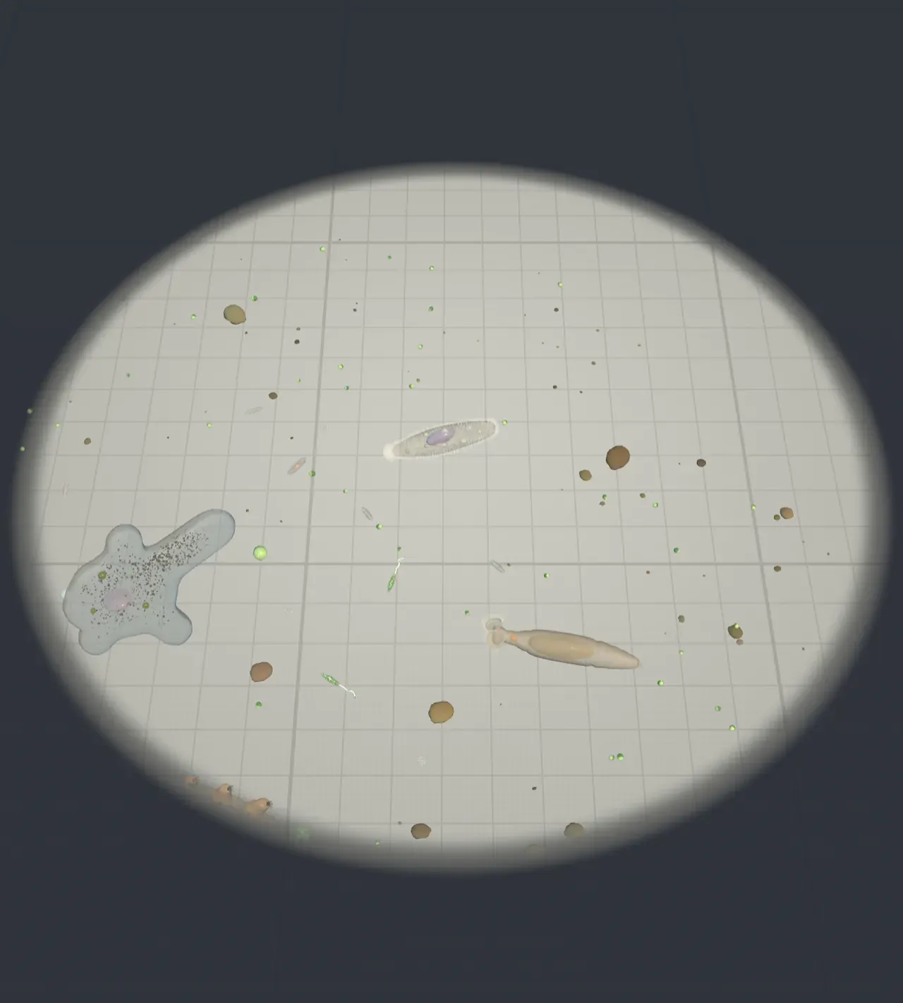
The Scene puts six of them in one drop: an amoeba, a paramecium, a euglena, a diatom, a rotifer and a tardigrade, with bacteria, small green algae, a filament of the alga Spirogyra and specks of debris around them. All are drawn at their true sizes relative to each other.
How the microscope shows them
At 10× the whole drop fits in view, a dome of water about 6 millimetres across, and everything in it is a speck. Turning up the magnification enlarges the drop around the organism you are following. A microscope does not let you see more of the drop as it magnifies; it shows you a smaller and smaller patch of it, enlarged to fill the same circle of light.
How big that patch is follows from the eyepiece. Inside it is a ring, the field stop, about 20 millimetres across, and the objective lens below enlarges the slide onto it. With an ordinary 10× eyepiece the patch of slide you see is about 200 millimetres divided by the total magnification: 5 mm at 40×, 2 mm at 100×, half a millimetre at 400× and a fifth of a millimetre at 1,000×. The chart beside the Scene marks that width on a ruler.
Try it: press the 10×, 40×, 100×, 400× and 1,000× presets under Magnification (powers of ten). Watch the ruled squares grow: at 1,000× one square of the finest grid, a hundredth of a millimetre, is wider than a bacterium is long.
Past about 1,000× a light microscope shows no new detail, because light itself is too coarse to resolve anything smaller than about a fifth of a micrometre. The Microscopes Article shows why, and how electron microscopes get past it.
Sizes side by side
A micrometre (µm) is a thousandth of a millimetre. In those units the drop holds:
- bacteria, about 2 µm long;
- a euglena, about 50 µm;
- a diatom, about 60 µm (most kinds are between 20 and 100);
- a paramecium, about 250 µm, a quarter of a millimetre;
- a rotifer, about 300 µm stretched out;
- a tardigrade, about 350 µm;
- an amoeba, about 500 µm, half a millimetre.
The unaided eye can just make out something a tenth of a millimetre across held at reading distance. So an amoeba, a paramecium or a tardigrade is a barely visible white dot in a jar held up to the light, and everything else is invisible.
The surprise is the number of cells. The amoeba, the paramecium, the euglena and the diatom are each one cell. The rotifer and the tardigrade are animals with a mouth, a gut, muscles and nerves, built from about a thousand cells each. Yet the single-celled amoeba is bigger than either of them. Size does not tell you how many cells something has.
Try it: set Follow to Amoeba and then Rotifer at 100×. The amoeba is one cell; the rotifer, smaller, is about a thousand.
One cell that does everything
The four single cells are not bacteria. Bacteria are far smaller and have no nucleus (see Bacteria). Each of these has a nucleus, mitochondria and other compartments wrapped in membranes, like the cells in your body (see Cells). Biologists call such single-celled eukaryotes protists. Each has to do on its own everything a whole animal does: find food, move, get rid of waste and keep its water balance.
They move in three different ways. The amoeba has no fixed shape. It pushes out lobes called pseudopods, “false feet”, and its runny inside streams forward into them while the back pulls in, so the whole cell flows across the surface at a few micrometres a second. The paramecium is covered in thousands of cilia, short hairs that beat in waves running down its body like wind over a field of wheat; they row it along at about a millimetre a second, several of its own lengths, spinning as it goes. The euglena has one long whip, a flagellum, that pulls it forward. The diatom has nothing that sticks out at all: it glides slowly by pushing a sticky mucus out through the raphe, a slit along its glass shell.
All four live in fresh water, which is a problem. A cell’s inside is saltier than pond water, so water keeps seeping in by osmosis, and a cell with no wall would swell and burst. The amoeba, the paramecium and the euglena each have a contractile vacuole, a bladder that fills with the extra water and squeezes it out every few seconds. The paramecium’s two are star-shaped, with canals that gather water from the whole cell.
Try it: follow the Paramecium at 400× and scrub Watch it feed a little way from Outside. Its body turns glassy, and you can watch its contractile vacuoles fill and empty in turn while the cilia ripple.
Hunters, grazers and gardeners
They also feed in different ways, which is what the end of Watch it feed shows.
The amoeba is a hunter. When it touches something it can eat, an alga or even a paramecium, two pseudopods flow out around it and meet on the far side, sealing it inside a bubble of membrane called a food vacuole, where enzymes digest it. This way of eating by engulfing is called phagocytosis, and your white blood cells use it to swallow bacteria.
The paramecium is a grazer. A funnel-shaped dip along its side, the oral groove, is lined with cilia that sweep a current of water and bacteria down to its cell mouth. The bacteria collect in a new food vacuole, which pinches off and is carried round the cell as it is digested; another forms every few minutes.
The euglena and the diatom are gardeners: they make their own food. The euglena’s green chloroplasts and the diatom’s gold-brown ones use sunlight to turn carbon dioxide and water into sugar, as plants do. The euglena stores what it makes as grains of a starch-like sugar, paramylon; the diatom stores droplets of oil. The euglena also has a red eyespot next to the base of its flagellum, which shades a light-sensitive spot on one side, so the cell can tell where the light is and swim towards it. In the dark it can take in food from the water instead.
Diatoms matter far beyond the drop. They live in every sea and lake, and together they make about a fifth of the oxygen produced on Earth each year. Their glass shells come in two halves that fit together like a box and its lid. When a diatom divides, each daughter keeps one half and builds a new, slightly smaller one inside it, so the population slowly shrinks until a round of sexual reproduction restores the full size.
Try it: follow the Euglena or the Diatom at 1,000× and scrub Watch it feed to Feeding. Their chloroplasts glow and their stored food appears grain by grain.
Animals smaller than a full stop
The rotifer and the tardigrade are animals, each with a brain, a gut and muscles, packed into less than half a millimetre.
The rotifer, or “wheel animal”, grips the glass with its toes and holds up its corona, two discs fringed with cilia. The cilia beat in a wave that runs round each disc, so they look like two spinning wheels. They draw a whirlpool of water towards its mouth, and inside its head a set of hard jaws, the mastax, grinds up the bacteria and algae it catches. Many rotifers keep exactly the same number of cells all their adult lives. The kind in the Scene, a bdelloid, is famous for another reason: no males have ever been found, and the females have been reproducing on their own for tens of millions of years. It can also dry out completely and revive when water returns.
The tardigrade, or “water bear”, lumbers about on eight stubby legs, each ending in hooked claws. It feeds by pushing two sharp stylets out of its mouth into a plant cell, such as one cell of the Spirogyra filament, and then pumping out the cell’s contents with its muscular pharynx. Tardigrades are the champions of survival: when their home dries up they pull in their legs, lose almost all their water and shrivel into a dry husk called a tun. In that state they have survived being frozen to near absolute zero, heated beyond boiling, and even exposed to the vacuum of space, and they wake up again when wetted.
Try it: follow the Tardigrade at 400× and scrub Watch it feed from Outside to Feeding. Watch the stylets reach into the alga cell, the green ribbon inside it shrink away, and the tardigrade’s gut turn green.
Who eats whom
Put together, the drop is a small food web. Bacteria feed on dead matter and dissolved food; the paramecium and the rotifer eat bacteria; the euglena, the diatom and the green algae make food from light; the amoeba eats algae and smaller protists; the tardigrade sucks out algae. Bigger animals in the pond, from water fleas to tadpoles and young fish, eat all of them. Almost all the life in a pond rests on things you cannot see.
What this model leaves out
The Scene draws each organism at its typical adult size, but real ones vary: an amoeba can be anything from a quarter to three quarters of a millimetre long. The six are placed far closer together than a real drop would usually hold them, so that they can be compared at 100×, and they swim, crawl and feed on the spot instead of wandering out of view, as real ones quickly do. The paramecium rocks rather than racing off at a millimetre a second.
Cilia and flagella beat many times more slowly than real ones so that their waves can be followed, and the paramecium carries about 1,500 cilia instead of several thousand. Colours are stronger than a real microscope shows: most protists are nearly clear and are seen mainly by their outlines and the granules inside them.
The microscope is simplified too. A real one focuses on a thin slice of the drop at a time, so things above and below it blur, and pond water is usually looked at under a thin glass cover slip, which flattens the drop. The Scene shows the whole depth sharp and leaves the drop uncovered. The field of view follows the usual 200 mm ÷ magnification for a 10× eyepiece; other eyepieces give slightly wider or narrower fields.