More magnification, no more detail
Scrape the inside of your cheek with a cotton bud, smear it on a glass slide, add a drop of stain and look through a school microscope. At 400 times you see flat, many-sided skin cells, each with a dark nucleus, and dotted over them some specks: bacteria from your mouth. Turn to the strongest lens, 1,000 times, and the specks become tiny rods and beads. On many of those bacteria sit viruses that infect them, but no light microscope will ever show you one, however much it magnifies.
The reason is the same one that stops you from enhancing a blurry photo on your phone. Spread two fingers and the picture grows, but past a point you only see bigger blurs. A microscope has two jobs: making things look bigger is the easy one, and making fine details come out separate is the hard one. The second is called resolution, and it is set by the light itself.
Two lenses, one after the other
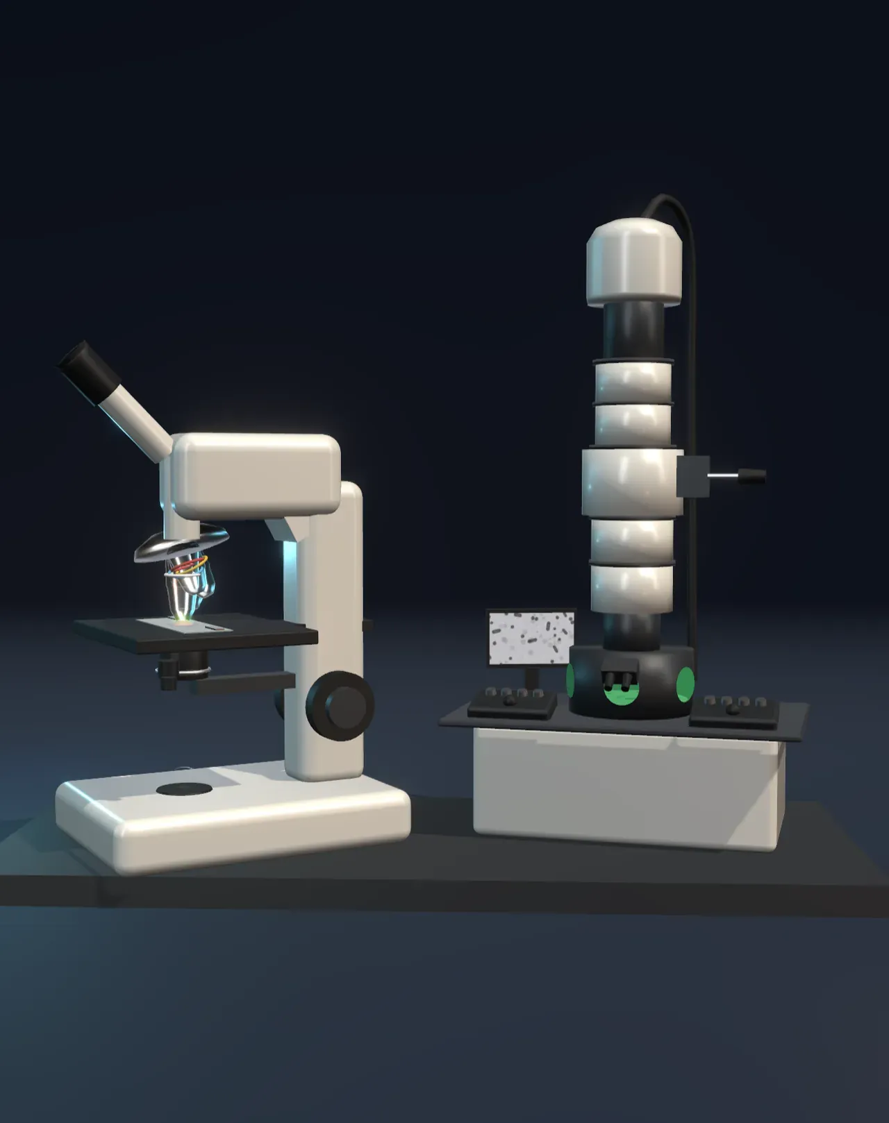
A light microscope magnifies in two steps. The objective lens, just above the specimen, gathers light from it and forms a real, enlarged, upside-down image inside the tube, 4, 10, 40 or 100 times the size of the specimen. The eyepiece then acts as a magnifying glass on that image and enlarges it 10 times more, so a 40× objective gives 400× in total. Below the stage, a condenser lens focuses the lamp’s light into a bright spot on the specimen. A turret holds several objectives, and the strongest works with a drop of oil between it and the slide. How a lens forms an image is in the Lenses Article; a telescope uses the same objective and eyepiece for things far away instead of close up.
Try it: choose Beam paths under Trace the beams. Follow the light up from the lamp, through the condenser and the specimen, to the enlarged image the objective makes inside the tube, upside down, and out of the eyepiece. Change Magnification (powers of ten) and the turret swings the next objective into place.
Why light runs out
Light is a wave, and a wave squeezing past something tiny spreads out, as the Diffraction Article shows. The objective catches only a cone of that spreading light, so it can never rebuild a point of the specimen as a point. Each one becomes a small blurred disc, and two details closer together than about the width of that disc merge into one. In 1873 Ernst Abbe, working out how to design better microscopes for Carl Zeiss, showed that this limit depends on two things only: the wavelength of the light, and how wide a cone the objective can collect, called its numerical aperture. Lord Rayleigh’s version of the rule puts the smallest detail at 0.61 times the wavelength divided by the numerical aperture.
No lens can collect a cone wider than a half-sphere, and oil between the lens and the slide lifts the numerical aperture to about 1.4 at best, so the limit sits near half a wavelength. With the 1.25 oil objective in the Scene, that is about 200 nanometres in violet light and 270 in green. A bacterium, about 2,000 nanometres long, is well above it. A virus that infects bacteria has a head about 100 nanometres across, and a ribosome is a quarter of that. They are below the blur, and no magnification brings them back. Magnifying beyond about 1,000 times the numerical aperture, around 1,250× for the best objectives, is called empty magnification: the picture grows, but nothing new appears.
Try it: set Magnification (powers of ten) to 10,000×. The electron picture shows the bacterium’s coat, the grains inside it and the viruses standing on it; the light picture is a soft blur. Change Light’s wavelength from 700 nm, red, to 400 nm, violet: the light picture sharpens a little, never enough.
Electrons have a wavelength too
In 1924 Louis de Broglie proposed that electrons, like light, travel as waves, with a wavelength that shrinks as they speed up (the Wave–particle duality Article shows them making fringes). An electron accelerated through 100,000 volts has a wavelength of about 0.0037 nanometres, roughly 150,000 times shorter than green light. In 1931 Ernst Ruska and Max Knoll built the first electron microscope in Berlin, and within two years Ruska’s instrument beat the light microscope. Ruska shared the 1986 Nobel Prize in Physics for it, and in 1939 the tobacco mosaic virus became one of the first viruses anyone had seen.
Glass would stop electrons, so an electron microscope focuses them with magnetic lenses: coils of copper wire in iron casings. A magnetic field pushes sideways on a moving electron, and the field of a coil bends electrons that pass off-centre back toward the middle, just as a glass lens bends light. Condenser coils shape the beam onto the specimen, an objective coil forms the first enlarged image, and further coils enlarge it again onto a fluorescent screen that glows where electrons land, or onto a camera. The whole column holds a vacuum, because air would scatter the beam.
Try it: at Beam paths, follow the electrons from the gun at the top through the coils. Both microscopes do the same job: one lens makes an enlarged image, the next enlarges that image again.
The price of seeing with electrons
Magnetic lenses are far from perfect. To keep their blur down, the beam must stay within a very narrow cone, and so an electron microscope resolves not a thousandth of a nanometre, as its wavelength alone would allow, but about 0.1 to 0.2 nanometres, roughly the size of an atom. That is still a thousand times finer than light.
The specimen pays for it. Electrons barely get through matter, so the specimen must be cut into slices thinner than about 100 nanometres, dried, stained with heavy metals such as uranium and lead to give contrast, and put in a vacuum: nothing in it is alive. The picture has no colour either, since electrons have none. The colours in published electron micrographs are painted on afterwards. A scanning electron microscope sweeps a fine beam over the surface of a specimen instead of shining through it, and gives pictures with a striking look of depth.
The light microscope keeps what electrons cannot: living cells, swimming and dividing, in colour. Since the 2000s, fluorescence microscopes have even slipped under the light limit by switching glowing molecules on a few at a time and locating each one precisely, work that won the 2014 Nobel Prize in Chemistry.
What this model leaves out
- Scale and setting. The electron microscope is drawn far smaller than life, and both are drawn without the operator. The specimen is stylised: a real cheek scraping would be too thick for electrons, and its skin cells, bacteria and viruses are placed for the view, not traced from a micrograph.
- The blur. Each picture is blurred by a smooth spot as wide as Rayleigh’s limit for its beam. A real image has faint rings round each point, and its contrast depends on the condenser’s setting and the stain. Light microscopes actually stop at about 1,000 to 1,500×; beyond that the light picture here is enlarged as a camera would enlarge it.
- The electron limit. It is worked out from the wavelength and a fixed aperture of 10 milliradians. In a real instrument the lenses’ aberrations, vibration and damage to the specimen set it, and correctors for the aberrations reach about 0.05 nanometres.
- The ray paths are drawn through single thin lenses. A real objective has many glass elements, modern microscopes add a tube lens, and electrons spiral through a magnetic lens, turning the image, rather than bending at one plane.
- One colour of light. The light is filtered to a single wavelength; white light behaves like a mixture, its blue end resolving finest.