A chain that folds itself
A protein is a chain of amino acids, often a few hundred long, joined end to end in an order set by a gene. How a cell reads that order out of its DNA and strings the chain together is told in From DNA to protein. This Article picks up where that one leaves off, with a finished chain floating in the water of a cell.
On its own a chain does nothing useful. A protein works because of its shape: a pocket that holds a sugar while it is cut, a groove that grips DNA, a channel just wide enough for one kind of ion. Within a fraction of a second of being made, most chains fold into that shape by themselves, and every copy of the same chain folds into the same one. Nothing tells the chain how. The instructions are in its own sequence.
Oil and water
Each amino acid has the same backbone, the part that links into the chain, and a side chain that sticks out from it. There are twenty kinds of side chain. Some are oily: they are mostly carbon and hydrogen, like the molecules of cooking oil. Others are water-loving: they carry an electric charge, or oxygen and nitrogen atoms that water can form hydrogen bonds with.
Water molecules cling to each other by hydrogen bonds, constantly breaking them and making new ones. An oily side chain cannot join in. The water next to it has to arrange itself round it in an orderly cage, which takes away some of the water’s freedom to tumble. When oily side chains crowd together, out of the water’s way, those cages are freed, the water gains disorder, and the whole system goes downhill. This pull is the hydrophobic effect, the same thing that gathers drops of oil on soup into larger drops. It is the main force that folds a protein.
So the first thing a fresh chain does is collapse. Within microseconds it crumples into a loose ball with most of its oily side chains on the inside, a state called a molten globule. It is compact but not yet tidy.
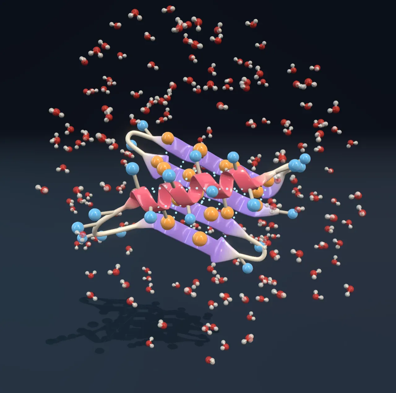
Try it: drag Let it fold to Fresh chain. Amber and blue beads are mixed all along the loose chain. Now move it slowly towards Settled chain and watch the amber beads gather in the middle while the blue ones stay outside, in the water.
Helices and sheets
Inside the collapsed ball the backbone has a problem. Every amino acid in it has an N–H group and a C=O group that made hydrogen bonds with water while the chain was loose. Buried, they must find partners in the chain itself, and there are two neat ways to pair them all up.
In an alpha helix the backbone winds into a spiral with 3.6 amino acids per turn, and each C=O bonds to the N–H four places further along. In a beta sheet stretches of the chain lie side by side, running up and down, and bond across to their neighbours like rungs of a ladder. Both were predicted from the shapes of the bonds by Linus Pauling and his colleagues in 1951, before anyone had seen a protein’s structure.
The protein in the Scene has one helix packed across a sheet of four strands. Along the helix the oily side chains form a stripe on the side that faces the sheet, and in the sheet every other side chain points towards the helix, so the oily ones from both meet in a tight core between them. Real proteins show this pattern again and again: oily and water-loving amino acids alternate along a strand and repeat every three or four along a helix.
Try it: stop Let it fold about three-quarters of the way along. The coral helix and lavender strands are taking shape, and the dotted hydrogen bonds are appearing between the strands and along the helix.
A narrow margin
A folded protein is not held together very strongly. Folding wins because burying oil frees water and the chain makes many weak bonds, but it loses the freedom of a loose chain to wriggle into countless shapes. The two nearly cancel. A typical small protein is stable by a few tens of kilojoules per mole, about as much as a handful of hydrogen bonds, out of hundreds of kilojoules on each side of the balance.
Heat tips that balance. The warmer the water, the more the disorder of a loose chain counts, until at the melting temperature half the molecules are folded and half are unfolded. Because a small protein folds and unfolds as one piece, the change is sharp: within ten degrees either side, almost all of them are folded or almost all unfolded. The chart shows this melting curve for the protein in the Scene, which melts at 60 °C.
Try it: with Let it fold at Settled chain, drag Temperature up from Body. Nothing much changes until about 50 °C. Then the helix and sheet loosen, and by 70 °C the chain is a loose, wriggling string with its oily side chains out in the water.
Unfolding a protein is called denaturing it. An unfolded enzyme stops working, which is why the Enzymes Article finds a peak in their activity just above body temperature. In this test-tube picture, cooling lets the chain fold again: around 1960 Christian Anfinsen unfolded the enzyme ribonuclease with chemicals, let it recover, and found it folded back into a working enzyme. That experiment showed that the sequence alone holds the shape.
Cooking is different. When an egg white is heated, its proteins unfold, and their exposed oily side chains stick to those of other unfolded chains. The proteins tangle into a mesh that turns the clear white solid and white. That is called aggregation, and it cannot be undone by cooling.
One change, or a hotter home
Because the margin is narrow, a small change can matter. Choose Mutant under Protein: one oily amino acid in the middle of the core has been swapped for a charged one, marked with a red ring. A charge buried away from water costs a lot, so the fold is weaker and melts at 45 °C. At body temperature about one molecule in ten is unfolded at any moment, and a fever leaves a quarter unfolded. Many inherited diseases work like this: the protein is made, but it is too unstable to stay folded, and the cell clears it away.
Microbes that live in hot springs have the opposite problem. Choose Heat-loving: this version keeps the same fold but has pairs of opposite charges on its surface, called salt bridges, which clip parts of the chain together. It melts at 90 °C. Proteins from heat-loving microbes really do have more salt bridges, tighter cores and shorter loops than ours; one of them, the copying enzyme of the hot-spring bacterium Thermus aquaticus, is used in PCR, the lab method for copying DNA, because it survives being heated nearly to boiling over and over.
Try it: choose Heat-loving and drag Temperature to 80 °C. The natural protein would have unfolded long before; this one is still folded, and gives way only near Boiling. Compare the curves on the chart: the same fold survives thirty degrees more heat.
Finding the shape in time
A chain of a hundred amino acids could take an astronomical number of shapes. Trying them one by one, even a trillion every second, would take far longer than the age of the universe, yet real proteins fold in microseconds to seconds. This puzzle is called Levinthal’s paradox. The answer is that folding is not a blind search. Every step that buries a little more oil or makes another hydrogen bond goes downhill, so the chain slides down a funnel of ever-better shapes towards the one at the bottom.
Not every chain makes it alone. Cells keep helper proteins called chaperones, which give a chain a sheltered place to fold and pull apart ones that start to clump. When folding goes wrong anyway, misfolded proteins can stack into stiff fibres called amyloid, which build up in the brain in Alzheimer’s disease. Predicting a protein’s shape from its sequence was one of biology’s great unsolved problems until about 2020, when computer programs trained on known structures learned to do it for most proteins.
What this model leaves out
The protein is made up, with one helix and four strands built from ideal shapes, though its fold is a common one in nature. Each side chain is drawn as one bead on a stick, oily or water-loving, where real side chains are clusters of atoms of twenty different shapes and sizes, and the backbone is drawn as a ribbon instead of its atoms. Water is shown as a sparse scatter of molecules, and in reality it fills every gap.
The Scene draws one molecule, but near the melting point a real protein flips between fully folded and fully unfolded, spending more or less time in each. The Scene shows that mix as a half-collapsed chain; the chart gives the true share. The folding path, from loose coil through a collapsed globule to the folded shape, is one typical route; real chains take many routes down the funnel. The melting curves come from a standard two-state model with typical values for a small protein, not measurements of a real one. Some proteins also unfold in the cold, far below 0 °C, which the model leaves out.