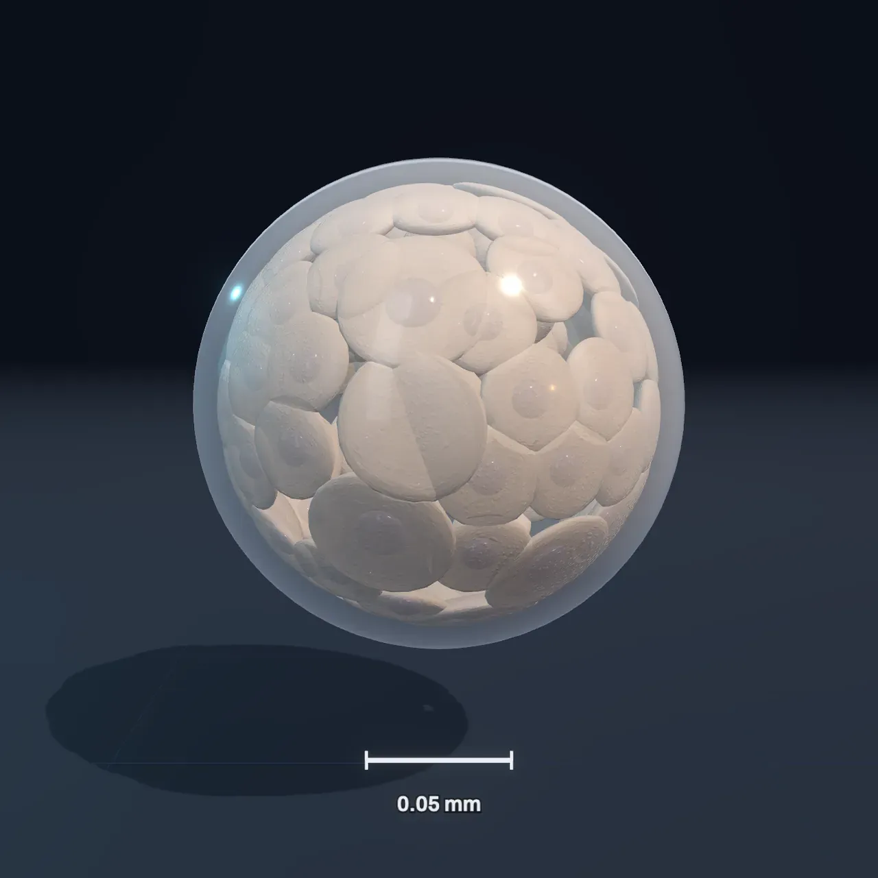
One cell, then many
A human life starts as one cell: an egg about a tenth of a millimetre across, fertilised by a sperm in the tube that leads to the womb. The fertilised egg, or zygote, sits inside a clear, tough shell called the zona pellucida. For its first hours it holds two small nuclei, one from the egg and one from the sperm, each with one set of chromosomes.
About a day after fertilisation it divides in two, then into four, then eight, each time by ordinary cell division. These first divisions are called cleavage, and they are unusual in one way: the cells do not grow between divisions. Each division halves the cells, so after four days of dividing the embryo is no bigger than the egg was. By day 4 it is a tight ball of 16 to 32 cells, the morula, named after the Latin for a mulberry.
Try it: move Time since fertilisation from Fertilised egg to Morula. The scale bar stays at 0.05 mm while the cells split into smaller and smaller pieces.
The cells of these first few days are totipotent: each could still make every part of a pregnancy, the body and the placenta too. That is why an early embryo that splits in two can grow into identical twins, two people with the same DNA.
A hollow ball
On day 4 and 5 the cells make their first choice. Cells on the outside of the ball flatten into a thin shell and pump fluid inward, opening a cavity; cells left inside cluster against one side. The embryo is now a blastocyst: a hollow ball of about a hundred cells, roughly a fifth of a millimetre across.
The outer shell is the trophoblast, and it will build the placenta, never the baby. The cluster inside, the inner cell mass, makes the whole body. Its cells are pluripotent: each can still become any kind of cell in the body, but no longer a placenta. Around day 6 the blastocyst squeezes out of its shell, and on day 7 it begins to sink into the lining of the womb. This is implantation.
Try it: press Blastocyst and choose Cut open, by fate under Cut open. The cut shows the ball is hollow, with teal cells round the outside and violet cells inside. Move Time since fertilisation on towards day 7 and the violet cells facing the cavity turn orange: they will line the yolk sac.
Three layers
In the second week the inner cell mass flattens into a disc of two layers. The upper layer, the epiblast, makes the whole body. The lower layer, the hypoblast, lines a bubble beneath the disc called the yolk sac; a second bubble, the amnion, forms above it and will one day hold the baby in its fluid.
Around day 15 a groove appears along the middle of the epiblast: the primitive streak. It marks where the head and tail will be and which side is left and which right. Cells of the epiblast crowd towards it and move in through it. Some spread out between the two layers and become the mesoderm; others push out the hypoblast and become the endoderm. The cells that stay on top are the ectoderm. This reshaping into three germ layers is called gastrulation, and each layer can now make only its own family of cells. Cells that can make several related kinds, but not all, are called multipotent.
| Germ layer | It makes, among others |
|---|---|
| Ectoderm | skin and hair, the brain, spinal cord and nerves, the lens of the eye |
| Mesoderm | muscle, bone, cartilage, the heart, blood and blood vessels, kidneys |
| Endoderm | the lining of the gut and lungs, the liver, the pancreas |
Try it: press Three layers and cut it open. Blue ectoderm is on top, a red layer of mesoderm spreads out from the streak, and yellow endoderm replaces the orange hypoblast underneath. The small cells drifting between the layers are moving in through the streak.
From a disc to a body
In the third and fourth weeks the flat disc turns into a body. Along its midline the ectoderm thickens into the neural plate, whose edges rise as two ridges and fold towards each other. They meet first at the future neck, around day 22, and close like a zip towards each end, making the neural tube: its front end will become the brain, and the rest the spinal cord. The head end closes around day 25 and the tail end a couple of days later. If part of the tube fails to close, a baby is born with a neural tube defect such as spina bifida; taking folic acid before and in early pregnancy makes these much rarer.
Beside the tube, the mesoderm breaks into pairs of blocks called somites, the first around day 20 and then about three a day, head end first. They will become the vertebrae, the muscles of the back and the deep layer of the skin. A tube of mesoderm under the head becomes the heart, which starts to beat around day 22, long before the embryo is the size of a grain of rice.
At the same time the whole disc folds. Its edges roll down and meet underneath, so the ectoderm wraps round the outside as skin and the endoderm is rolled into a tube inside, the gut. Head and tail curl down as well, and the heart swings round under the head. By four weeks the embryo is a curled body about 4 or 5 mm long, with a large head, ridges in the neck that will form the jaw and throat, a bulging heart, a tail, and small buds that will grow into arms and legs.
Try it: press Neural tube and watch the folds zip shut in the middle while both ends are still open. Then move on to Body plan and cut it open: the blue neural tube runs along the back, the yellow gut along the front, and the red heart sits under the head.
How cells specialise
Every cell of the embryo carries the same DNA. What makes a nerve cell different from a muscle cell is which of its genes are switched on, and so which proteins it makes (from DNA to protein). A cell decides by listening to its neighbours. Signalling molecules spread from groups of cells such as those at the streak, and a cell reads how much of each signal reaches it to tell where it is. In reply it switches on transcription factors, proteins that turn other genes on and off and often keep themselves switched on.
Each decision narrows the next. A fertilised egg can make anything; an inner cell can make any part of the body; a mesoderm cell only muscle, bone, blood and their relatives; and in the end a cell is specialised, or differentiated, as one of more than 200 kinds. The choices are mostly one-way and are passed on at every division, which is why a skin cell divides into skin cells and not into liver. The chart beside the Scene shows this narrowing as a family tree, coloured as the Scene colours the cells when you cut it open.
Stem cells
A stem cell is a cell that can both copy itself and make more specialised cells. The pluripotent cells of the inner cell mass are the first stem cells, and they last only a few days in the embryo. Grown in a dish, they become embryonic stem cells, which can divide indefinitely and still make any kind of cell.
Adults have stem cells too, but they are multipotent: each kind keeps one tissue going. Stem cells in the bone marrow make every kind of blood cell, which is why a bone-marrow transplant can rebuild the blood of a person with leukaemia; stem cells deep in the lining of the gut replace it every few days, and when their controls fail they can give rise to cancer. In 2006 researchers found that switching on four genes can turn a mature skin cell, first a mouse’s and a year later a person’s, back into a pluripotent one: an induced pluripotent stem cell, a discovery recognised with a Nobel Prize in 2012.
Stem cells are used to study how diseases develop and to test drugs, and blood stem cell transplants are standard treatment; therapies with cells grown from pluripotent stem cells, for the eye, the heart or the brain, are being tested in trials. Research on human embryos is tightly regulated, and many countries allow it only up to 14 days after fertilisation, about when the primitive streak appears and an embryo can no longer split into twins.
What this model leaves out
The Scene follows typical human timings, rounded, and every embryo runs a little early or late. It draws each early cell, up to 256, dividing at about the right times. From the second week it draws the embryo as smooth tissue printed with cells of about the right size, which shrink on the screen as the embryo grows. It leaves out the journey down the tube to the womb, the womb itself, the placenta and the outer membranes, the connecting stalk that becomes the umbilical cord, and the cells that will make eggs or sperm. The amnion is shown only while the disc is flat, though in life it grows to surround the whole embryo.
The colours show fates, not anything a microscope would see, and fates are less sharp than the colours suggest: a cell’s future is settled gradually, by the signals around it, and some cells keep more options than their layer suggests. The folding is simplified to a sheet that rolls and curls, and the heart is drawn as a simple looped tube. For how one cell divides, see cell division; for what is inside each cell, see cells.