A lining that renews itself
The lining of your gut is replaced every few days. Deep in the lining, a few stem cells keep dividing; their daughters move up, do their job of absorbing food and making mucus, and are shed. Across the whole body, hundreds of billions of cells are replaced every day. Each new cell comes from cell division, and each division copies about six billion letters of DNA.
Almost all of this goes right. The tissue stays the same thickness for a lifetime because every cell obeys a set of controls: it divides only when its neighbours signal that a cell is needed, it stops when told to, and it checks its DNA before and during each division. The Scene starts with Mutations at 3, partway along the path this Article follows.
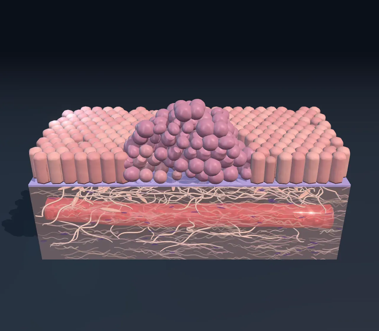
Try it: set Mutations to Healthy. The lining is one even layer of tall cells on a thin sheet, the basement membrane, with the soft connective tissue and a blood vessel beneath. Nothing grows out of place.
Brakes, accelerators and checkpoints
A cell’s controls are proteins, and each protein is built from a gene. Two kinds of gene matter most. Some act like an accelerator: they pass on a signal to divide, and a mutation that leaves one stuck on makes the cell divide without being asked. These are called oncogenes when mutated; a common example is KRAS. Others act like a brake: they stop division or hold the cell back. These are tumour-suppressor genes, such as APC. A cell has two copies of every gene, one from each parent, and one working copy of a brake is usually enough, so both copies must be broken before the brake fails.
Between the brakes and the accelerators sit the checkpoints: points in the cycle of growing, copying its DNA and dividing where the cell stops and checks itself. If the DNA is damaged, a protein called p53 halts the cycle while the damage is repaired, and if it cannot be repaired, it makes the cell destroy itself, a tidy death called apoptosis. Because it guards against so much, p53 is the most often broken gene in cancer: its gene, TP53, is mutated in about half of all tumours.
Try it: with Mutations at Polyp, choose On under Show the checkpoints. The cells turn to glass and their nuclei show what each is doing. Many cells in the polyp are dividing, and many of those have damaged their DNA on the way, but they are caught: amber cells are held at a checkpoint, and brown fragments are cells that have destroyed themselves. Now add a third mutation. The amber cells are gone, and red ones divide with their damage.
One mutation is not enough
Every division leaves a few new mistakes in the copied DNA, and sunlight, tobacco smoke and other chemicals add more. Most mutations land where they do no harm. One that breaks a control helps its cell divide a little more than its neighbours, and its descendants, a clone, slowly spread. Among them, another mutation breaks a second control, and that cell’s descendants outgrow the first clone. The Scene shows this as cells that grow darker with each mutation they carry. It is natural selection inside the body: cells that divide faster leave more descendants, whether or not that is good for the body.
This is why cancer usually needs several mutations in one lineage, why it takes years or decades, and why it mostly affects older people. It is also why some families have more cancer: a person can inherit one broken copy of a brake gene, such as BRCA1, BRCA2 or APC, so their cells start one step along the way.
Try it: step Mutations from 0 to 4 and watch the chart beside the Scene. One mutation makes a crowded patch of about ten thousand cells, too small to see. Two make a benign polyp. With three, the lump becomes unstable: its cells vary in size, their nuclei are large and dark, and new mutations arise faster because nothing stops a damaged cell.
From a lump to a cancer
A lump of cells needs food and oxygen, which seep only a millimetre or two from the nearest blood vessel. To grow further it must call in new vessels, and at Lump you can see them sprouting from the vessel below and winding up into it. With its own blood supply a lump can reach a centimetre, about a billion cells, the size at which it may be felt or seen on a scan (medical imaging).
So far the lump is a benign tumour: however large, its cells stay on their side of the basement membrane. The next step is the one that makes it cancer. At 5 mutations the cells cut through the membrane and push into the connective tissue in cords and single cells. A tumour that invades this way is called malignant. Most cancers start in linings like this one, in the skin, gut, lungs, breast or prostate, and are called carcinomas; others start in blood cells, bone or other tissues.
Try it: switch Mutations between 4 and 5 with Show the checkpoints at Off. From above the lump looks much the same. Only the cut shows whether the membrane has been broken, which is why doctors examine a thin slice of a tumour under a microscope to tell benign from malignant.
Spreading
At 6 mutations, cells that have reached the blood vessel squeeze through its wall and are carried away. Most die in the blood, but a very few settle in another organ, such as the liver, lungs or bones, and grow into a new tumour there: a metastasis. Cancers can also spread through the lymph vessels to nearby lymph nodes. Most deaths from cancer are caused by tumours that have spread, which is why finding a cancer before it can spread matters so much.
Prevention, screening and treatment
Many cancers can be prevented or caught early. Not smoking avoids most lung cancers; protection from strong sunlight lowers the risk of skin cancers; and the vaccine against human papillomavirus prevents most cancers of the cervix, a reminder that a few viruses can cause cancer too. Screening looks for the early steps: a camera test of the bowel can find and remove polyps before they become cancer, and a cervical test can find abnormal cells years before they invade.
Treatment depends on how far the steps have gone. A tumour that has not spread can often be removed by surgery, sometimes with radiotherapy. Drugs that travel through the whole body reach cancer that has spread: some kill dividing cells, many by jamming the spindle that pulls chromosomes apart; some block the exact protein a mutation left switched on; and some help the immune system, which already destroys many abnormal cells, to recognise the tumour. Those drugs are called immune checkpoint inhibitors, but the checkpoints they release are brakes on immune cells, not the checkpoints of the cell cycle. Survival has risen for many cancers, and in many countries most people diagnosed with several common ones now live for many years afterwards.
What this model leaves out
The Scene runs six mutations in one fixed order, each switching off one control. Real cancers carry a handful of such driver mutations among thousands of harmless passenger mutations, in orders that differ from tumour to tumour and from one kind of cancer to another; some controls are lost by switching genes off without changing their letters. The order here follows a common path for bowel cancer, and the genes named on the chart are examples, not the only ones.
The lump is drawn a few hundred cells big, and every step is shown at once; in a person each step takes months to years, and a real lump grows to millions or billions of cells. The shares of cells resting, dividing or held at checkpoints are illustrative, and the colours are for teaching: under a microscope, stains show nuclei purple and the rest of the cell pink. The Scene leaves out the immune cells that patrol the tissue, the support cells that tumours recruit, and the lymph vessels. For how a single cell divides, see cell division; for how genes can be edited to study or treat cancer, see CRISPR and gene editing.