One job, many senses
Your eyes, ears, nose, tongue and skin look nothing alike, yet they all do the same job. Each holds receptor cells, and each kind of receptor is built to respond to one kind of stimulus: light, a push, a molecule, heat. When its stimulus arrives, the receptor’s membrane lets ions through, and the voltage across it shifts. That shift, the receptor potential, is bigger for a stronger stimulus. Past a threshold it makes the nerve fibre fire spikes, the brief all-or-nothing pulses described in the nervous system Article. This step, from a stimulus to an electrical signal, is called transduction, and it is where every sense begins.
The rest is the same for every sense. Spikes are the only signal a nerve fibre can send, and one spike looks like any other. A fibre reports how strong its stimulus is by how often it fires, and what kind of stimulus it was by which fibre it is. In the 1830s the physiologist Johannes Müller noticed that it does not matter how a nerve is set off: press gently on a closed eye and you see light, because the optic nerve can only ever report light. The brain reads its senses by which wires carry the spikes, and it sorts them into sight, sound and touch in its own areas, shown in The brain.
Vision and Hearing follow light to the rods and cones and sound to the hair cells of the cochlea. This Article starts with the skin, which holds several senses in one place.
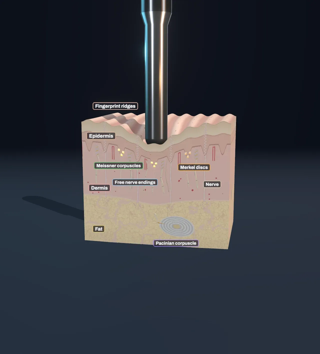
Try it: leave the Scene as it starts and watch the probe come down. The amber Merkel discs under it flash for as long as it presses; the green and violet corpuscles flash only as it goes in and comes out.
Four ways to feel a touch
The pad of a fingertip is one of the most sensitive patches of skin you have. Each square centimetre holds the endings of about 240 touch fibres, and two points pressed on it about 2 mm apart already feel like two. On your back they need to be several centimetres apart. Under the fingerprint’s ridges sit four main kinds of touch receptor, each the ending of one nerve fibre wrapped in its own structure:
- Merkel discs are small cells at the base of the epidermis, the outer layer of skin, each pressed against a disc-shaped nerve ending. They sit under the fingerprint’s ridges and feel where the skin is pressed and how hard: edges, corners, the dots of Braille.
- Meissner corpuscles are oval stacks of flattened cells with the nerve ending zigzagging between them, tucked just under the epidermis between the ridges. They feel the skin moving: an object starting to slip in your grip, or a flutter of taps.
- Pacinian corpuscles are about a millimetre long, big enough to see without a microscope, and sit deep in the fat beneath the skin. Up to seventy thin layers wrap the nerve ending like an onion. They feel vibration, through a tool or a table as well as at the skin itself.
- Ruffini endings, spindles that feel the skin stretching, are a fourth kind, though how many there are in a fingertip is still argued over. The Scene leaves them out.
In each of them the nerve ending’s membrane holds ion channels that open when it is stretched. The main one, Piezo2, was identified only in 2010. In 2021 Ardem Patapoutian shared the Nobel Prize for finding it, with David Julius for the heat sensor that comes up below.
Try it: drag Press from 0.3 mm to 1.5 mm. The dent in the skin deepens and the Merkel discs fire faster, from about 37 spikes a second while a light press is held to about 66 for a hard one. Then slide it down to 0 mm: the probe rests on the skin without pressing in, and the touch receptors stay quiet.
Fast and slow
If the four receptors only reported pressure, they would be redundant. What separates them is adaptation: how long a receptor keeps firing while its stimulus stays the same.
Merkel discs are slowly adapting. They fire hard as the skin is pressed in and then settle to a steady rate for as long as the press lasts. Meissner and Pacinian corpuscles are rapidly adapting: they fire while the skin is moving and fall silent as soon as it stops, then fire again when it is let go. That is why you feel your clothes as you put them on and then stop noticing them. The Pacinian corpuscle’s layers are part of the reason. Squeezed slowly, the fluid between them flows aside and the steady pressure never reaches the nerve ending; only a quick change gets through.
Fast receptors are also tuned to how fast the skin vibrates. Meissner corpuscles follow slow flutter best, around 30 times a second, with one spike for each tap. Pacinian corpuscles follow fast vibration best, around 250 times a second, and at that rate they need the skin to move less than a micrometre, a thousandth of a millimetre. A phone buzzing in your pocket, the texture of a surface you drag a pen across and the rumble of a train through the floor all reach you mostly through them.
Try it: set Press to 0.8 mm and slide Vibration from 0 Hz to 30 Hz, 80 Hz and 250 Hz. At 30 Hz only the Meissner corpuscles keep time, at 80 Hz both kinds do, and at 250 Hz only the Pacinian corpuscle, deep down, fires on every cycle. The Merkel discs keep reporting the press throughout.
Warm, cold and pain
Temperature and pain have no special structure around their receptors. They are free nerve endings: bare branches of thin fibres that reach up into the living layers of the epidermis. What makes each one specific is a channel in its membrane that opens at a temperature.
- Cold endings carry TRPM8, which opens below about 26 °C, and also when menthol binds to it. That is why mint feels cool.
- Warm endings fire more as the skin warms from about 32 °C to about 43 °C, and stop above that.
- Pain endings, or nociceptors, carry TRPV1, which opens above about 43 °C and also when capsaicin, the hot substance in chilli, binds to it. Others respond to extreme cold, to crushing, or to chemicals released by damaged tissue.
Skin at rest is at about 33 °C, and the free endings tick over slowly there. Like the touch receptors, they report change best: step into a hot bath and it feels hotter at first than a minute later, though the water has barely cooled.
They are also slower. Touch travels in thick fibres wrapped in myelin, at about 50 metres a second. Cold travels in thinner myelinated fibres, and warmth and most pain in thin, bare C fibres at about a metre a second. That is why stubbing a toe hurts twice: a sharp first pain carried by faster fibres, and a dull ache that arrives a second or so later.
Try it: choose Nerve signals under Show the nerve signals, then slide Probe temperature to 5 °C and then to 48 °C. At 5 °C the cold endings and the pain endings both fire; at 48 °C the warm and pain endings fire, and their beads crawl down their thin fibres far behind the touch spikes.
Smell and taste
Smell and taste are chemical senses: their receptors respond to molecules.
In the roof of each nostril is a patch of lining the size of a postage stamp, with millions of olfactory receptor neurons. Each sends fine hairs into the mucus, and each makes just one kind of receptor protein out of roughly 400 that human genes encode. A molecule that fits sets off a chain of reactions inside the cell, which opens ion channels and fires spikes straight to the olfactory bulb under the front of the brain. Most smells set off several kinds of receptor at once, and the brain recognises the combination, which is how 400 kinds of receptor can tell thousands of smells apart. Linda Buck and Richard Axel won a Nobel Prize in 2004 for finding the receptor genes. Olfactory neurons are among the few nerve cells that are replaced throughout life.
Taste uses taste buds, clusters of 50 to 100 cells in the bumps on the tongue and the back of the mouth. Different cells respond to sweet, umami (the savoury taste of glutamate), bitter, salty and sour, the first three through receptor proteins and the last two through ions passing straight through channels. Most of what we call the taste of food is really smell, carried up from the back of the mouth into the nose, which is why food tastes flat when you have a cold.
Senses we lack, and senses beyond five
Five senses is a schoolbook number. Your inner ear also senses balance and acceleration, stretch receptors in muscles and tendons tell you where your limbs are with your eyes closed, and receptors inside the body report a full stomach, blood pressure and the carbon dioxide in your blood.
Other animals use receptors tuned to stimuli we cannot sense at all, and the job is the same:
- Honeybees see ultraviolet light: their three kinds of light receptor cell respond best to ultraviolet, blue and green, where ours respond best to blue, green and red. Many flowers are patterned in ultraviolet, with marks that guide a bee to the nectar.
- Pit vipers, such as rattlesnakes, have a pit between eye and nostril holding a thin membrane full of free nerve endings with TRPA1, a channel that opens as it warms. Infrared from a mouse warms the membrane by a tiny fraction of a degree, and the snake strikes in the dark.
- Bats shout at pitches far above our hearing and listen to the echoes, building a picture of moths and branches from sound alone, with hair cells like ours tuned to higher pitches.
- Sharks sense the faint electric fields of a fish’s muscles through jelly-filled pores on their snouts, and many birds sense the direction of the Earth’s magnetic field, by a mechanism still not settled.
What this model leaves out
The Scene shows one patch of skin, 4 mm across, and one probe pressing on it. The press, its timing and the vibration’s size (20 micrometres) are fixed; only the depth, the frequency and the temperature change. Each kind of receptor follows a simple rule: a Merkel disc’s steady rate grows with the depth of the press and levels off, Meissner and Pacinian corpuscles fire only as the skin moves, and each kind follows vibration above a threshold curve with its lowest point at about 2, 30 and 250 Hz. The free endings follow typical curves of rate against skin temperature, warmed or cooled under the probe over a fraction of a second. Real receptors of one kind vary a great deal, and the numbers are typical values from recordings, not measurements of one hand.
A real fingertip holds many more receptors than the few dozen drawn, each of its touch fibres serving several endings over an area a few millimetres across, and the fibres themselves are thinner than drawn. Ruffini endings, hair follicles (there are none on a fingertip pad) and the blood vessels’ own nerves are left out, and so are the brain’s maps that turn the spikes into a feeling of where, how hard and how warm.