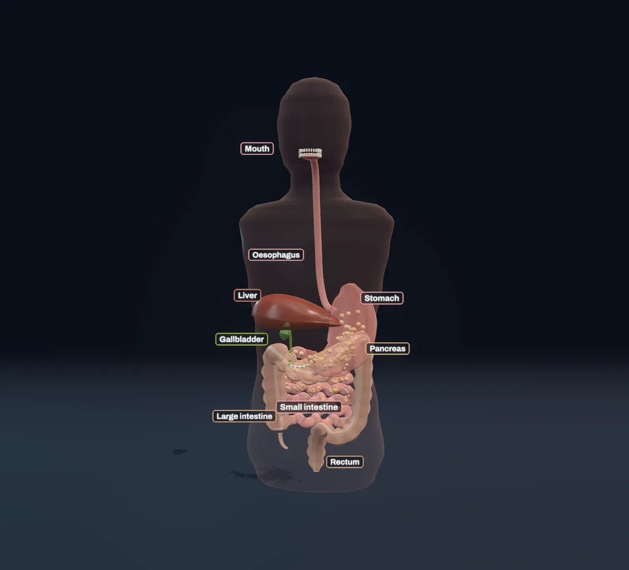
A tube through the body
From mouth to anus, your gut is one long tube, open at both ends. Food inside it is not really inside you yet: it is in a tunnel through you, much as the hole is not part of a doughnut. To use a meal, the body has to get it across the tube’s wall and into the blood, and that wall lets through only small molecules. Bread, meat and butter are made of very large ones: starch, proteins and fats. Digestion is the work of cutting them down to pieces small enough to cross.
The tube is the mouth, the oesophagus (gullet), the stomach, the small intestine and the large intestine, which ends in the rectum. Beside it sit three organs that pour juices into it: the salivary glands, the pancreas, and the liver with its gallbladder.
Try it: leave Meal on All three and slide Time since eating from 0 to 3 hours and then to 12, the next morning. Watch where the specks of food are, and how fast the ones in the small intestine shrink.
Mouth and stomach
Chewing breaks food into a paste with more surface for juices to work on. Saliva wets it, and its enzyme, amylase, starts cutting starch at once, which is why bread held in the mouth for a minute begins to taste sweet. A swallow then takes about eight seconds to reach the stomach, pushed down the oesophagus by a ring of squeezing muscle that travels along it.
The stomach is a muscular bag that stretches to hold a litre or more. Its lining pours out hydrochloric acid, strong enough to kill most germs that come in with the food, and pepsin, an enzyme that starts cutting proteins and works best in acid. Waves of squeezing sweep down its lower part about three times a minute, grinding the food against the closed outlet, the pylorus, until it is a thick soup called chyme. Only then does the pylorus let it through, a few millilitres at a time.
So the stomach is mostly a store and a mill. Very little is absorbed there; alcohol and some medicines are exceptions.
Try it: slide Time since eating from 0 to 1 hour. Most of the meal is still in the stomach, the specks turning soupy, with the next ones to leave crowded down by the outlet.
The small intestine does the work
Almost all digestion and absorption happen in the small intestine, a tube about as thick as a finger and several metres long, folded back and forth in the belly. Its first stretch, the duodenum, is where the chyme meets two juices. The pancreas sends enzymes for every kind of food, with bicarbonate that neutralises the stomach’s acid, so that the gut’s own enzymes, which work best in neutral water, can get going. The liver makes bile, which is stored and concentrated in the gallbladder and squeezed out when food arrives. Bile has no enzymes; its job is to break fat up.
Food takes about three to five hours to travel along the small intestine, pushed forward by slow waves and mixed by rings of muscle that tighten and relax in place. The specks shrink as it goes, most of them in the first metre or two.
Its inside is built for absorbing. The lining is folded into ridges, the ridges are covered with villi, fingers about a millimetre tall, and every cell on a villus wears thousands of tiny microvilli, a fuzz called the brush border. Together they give a surface of about thirty square metres, the floor of a large room, packed into a tube you could hold in two hands.
Cutting food down to size
Every large food molecule is a chain of smaller units, joined by bonds that water can split. Left alone, that would take years. Enzymes do it in a moment, each kind fitting only its own kind of bond:
- Starch is a chain of hundreds to thousands of glucose units. Amylase from saliva and the pancreas cuts it into two-glucose pieces, and enzymes on the brush border split those into single glucose.
- Proteins are chains of amino acids. Pepsin in the stomach, then trypsin and other enzymes from the pancreas, cut them into short pieces, and brush-border enzymes finish them off as single amino acids, or pairs and threes that the cells take in and split inside.
- Fat does not mix with water, so enzymes can reach only the surface of a big drop. Bile salts coat it and break it into droplets, which gives far more surface. Lipase from the pancreas then cuts each fat molecule into fatty acids and a leftover piece of glycerol.
Try it: choose Gut lining under Zoom to the gut lining and pick Rice (starch), then Chicken (protein), then Butter (fat). Each kind of food arrives from its own side; watch which enzyme meets it and what comes out.
Into the blood, or into the lymph
The cells of the villi take the pieces in. Glucose comes in with sodium through a carrier in the cell’s membrane, and is passed out the other side into the blood; amino acids use carriers of their own. The rest of the way is diffusion: each villus holds a net of blood capillaries just under its surface. The blood draining the gut does not go straight back to the heart. It collects into the portal vein and runs first through the liver, which stores much of the sugar, builds proteins from the amino acids and clears out anything harmful before the blood moves on to the rest of the circulation.
Fat takes a different road. Fatty acids slip through the cell membrane, and inside the cell they are rebuilt into fat and packed with protein into droplets called chylomicrons. These are too big to get into the capillaries, so they enter the lacteal, a lymph vessel up the middle of every villus. Lymph carries them up through the chest and pours them into a large vein near the neck, so most fat reaches the blood without passing through the liver first. After a fatty meal there are so many droplets that the lymph from the gut looks milky; that is where the word lacteal, “milky”, comes from.
Try it: with Zoom to the gut lining on Gut lining and Meal on All three, follow the beads. Gold sugars and coloured amino acids disappear into the red capillary and away along the vein; the fat comes out of the villus as a pale droplet sliding down the lacteal into the lymph vessel below.
Why fat stays longer
How fast the stomach empties is controlled from the duodenum. When fat and protein arrive there, its cells release hormones, cholecystokinin above all, that squeeze the gallbladder and the pancreas into action and hold back the pylorus, so the stomach lets food out more slowly. It makes sense: fat needs the most work in the small intestine, and the gut gives itself time to do it. A meal that sits longer in the stomach is also one reason you stay full for longer.
In the Scene the stomach takes about 1.3 hours to let out half a meal of rice, 1.8 hours for chicken and 2.8 hours for butter. The rice is mostly absorbed within about three hours, the fat only after six or seven.
Try it: set Time since eating to 3 hours and switch Meal between Rice (starch) and Butter (fat). The rice has almost all left the stomach; nearly half of the butter is still there. The chart shows the whole day for each.
The large intestine
What reaches the large intestine is what the small intestine could not use: mostly fibre from plants, with a few per cent of the starch, protein and fat, and a great deal of water. Each day about nine litres of fluid pass into the gut, from what you drink and from all the juices it makes; the small intestine takes back most of it, and the large intestine most of the rest, leaving the leftovers as firm stool.
The large intestine is wider, gathered into pouches, and slow: food spends one to three days in it. It is also home to trillions of bacteria that feed on the fibre we cannot digest and make useful substances from it. Those partners are a story of their own, told in The microbiome and symbiosis; what you choose to eat in the first place is the subject of Nutrition.
Try it: slide Time since eating to 12 hours, the next morning. The food has long been absorbed, and only a small brown remainder is still creeping round the large intestine.
What this model leaves out
The Scene shows one meal on an empty gut. Real meals overlap, and the time a meal takes varies widely: the numbers here are typical of a healthy adult and come from a simple model, not a measurement. The stomach lets the meal out along a smooth curve fitted to measurements of stomach emptying, each share of it then takes between two and a half and four and a half hours through the small intestine, and each nutrient is absorbed along it at a rate chosen to put most absorption in its first half. Real stomachs empty liquids faster than solids and sort what they let out by particle size; sugary drinks, fibre, meal size and exercise all change the timings.
Each meal stands for food made of one nutrient. Rice has some protein, chicken some fat, and nobody eats butter alone. The mixed meal treats its starch, protein and fat as leaving the stomach together. Water, vitamins and minerals, and the hormones that steer digestion are left out, and so are the gut bacteria.
The gut is drawn smaller and simpler than life: the small intestine is folded into a tube of under two metres where a real one is several metres long, and the large intestine’s bacteria are not shown. On the lining, villi are drawn hundreds of times larger than life and molecules about a million times larger, so that both show at once, and their motion is slowed down a thousand times. Real starch chains are hundreds of units long, not eight.